
Page 308 - Cardiac Nursing
P. 308
0:4
0:4
1
1
2 A
M
M
2 A
M
009
3
3
xd
3
0/0
6/2
009
0/0
6/2
p
p
A
p
t
ara
ara
t
ara
A
g
g
Pa
Pa
g
84
84
e 2
e 2
xd
13_
13_
0-c
p
p
LWB
K34
LWB
K34
LWBK340-c13_ pp277-290.qxd 30/06/2009 10:42 AM Page 284 Aptara
27
90.
q
90.
q
q
7-2
0-c
27
7-2
284 P A R T III / Assessment of Heart Disease
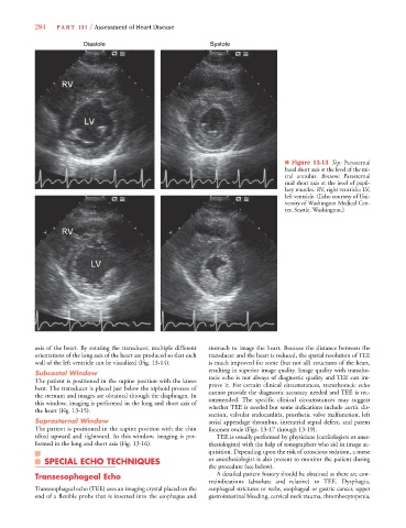
Diastole Systole
■ Figure 13-13 Top: Parasternal
basal short axis at the level of the mi-
tral annulus. Bottom: Parasternal
mid short axis at the level of papil-
lary muscles. RV, right ventricle; LV,
left ventricle. (Echo courtesy of Uni-
versity of Washington Medical Cen-
ter, Seattle, Washington.)
axis of the heart. By rotating the transducer, multiple different stomach to image the heart. Because the distance between the
orientations of the long axis of the heart are produced so that each transducer and the heart is reduced, the spatial resolution of TEE
wall of the left ventricle can be visualized (Fig. 13-14). is much improved for some (but not all) structures of the heart,
resulting in superior image quality. Image quality with transtho-
Subcostal Window
The patient is positioned in the supine position with the knees racic echo is not always of diagnostic quality and TEE can im-
bent. The transducer is placed just below the xiphoid process of prove it. For certain clinical circumstances, transthoracic echo
the sternum and images are obtained through the diaphragm. In cannot provide the diagnostic accuracy needed and TEE is rec-
this window, imaging is performed in the long and short axis of ommended. The specific clinical circumstances may suggest
the heart (Fig. 13-15). whether TEE is needed but some indications include aortic dis-
section, valvular endocarditis, prosthetic valve malfunction, left
Suprasternal Window atrial appendage thrombus, interatrial septal defect, and patent
The patient is positioned in the supine position with the chin foramen ovale (Figs. 13-17 through 13-19).
tilted upward and rightward. In this window, imaging is per- TEE is usually performed by physicians (cardiologists or anes-
formed in the long and short axis (Fig. 13-16). thesiologists) with the help of sonographers who aid in image ac-
quisition. Depending upon the risk of conscious sedation, a nurse
SPECIAL ECHO TECHNIQUES or anesthesiologist is also present to monitor the patient during
the procedure (see below).
Transesophageal Echo A detailed patient history should be obtained as there are con-
traindications (absolute and relative) to TEE. Dysphagia,
Transesophageal echo (TEE) uses an imaging crystal placed on the esophageal strictures or webs, esophageal or gastric cancer, upper
end of a flexible probe that is inserted into the esophagus and gastrointestinal bleeding, cervical neck trauma, thrombocytopenia,