Page 310 - Cardiac Nursing
P. 310

2 A
                                          M
                                      0:4
                                        2 A
                                            Pa
                                            Pa
                                          M
                                          M
                                      0:4
                                6/2
                                6/2
                              0/0
                              0/0
                                     1
                                     1
                                  009
                                  009
                                                   p
                                                    t
                                                   p
                                                   p
                                                     ara
                                                     ara
                                                    t
                                                     ara
                                                  A
                                              g
                                               e 2
                                              g
                                              g
                                                86
                                                  A
                                               e 2
                                                86
                   p
                    27
                 13_
                   p
                      7-2
                        90.
                    27
                      7-2
         LWB
            K34
         LWBK340-c13_ pp277-290.qxd  30/06/2009  10:42 AM  Page 286 Aptara
         LWB
               0-c
                 13_
            K34
               0-c
                              3
                           xd
                             3
                             3
                           xd
                          q
                        90.
                          q
                          q
                  286    P A R T  III / Assessment of Heart Disease
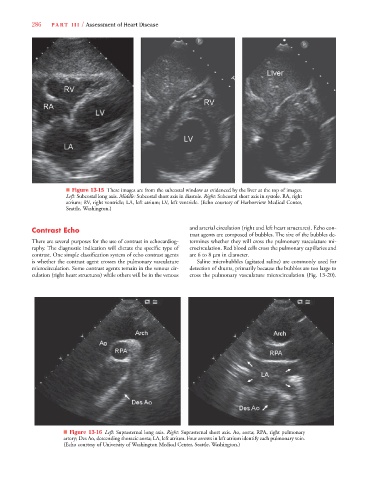
                              ■ Figure 13-15 These images are from the subcostal window as evidenced by the liver at the top of images.
                              Left: Subcostal long axis. Middle: Subcostal short axis in diastole. Right: Subcostal short axis in systole. RA, right
                              atrium; RV, right ventricle; LA, left atrium; LV, left ventricle. (Echo courtesy of Harborview Medical Center,
                              Seattle, Washington.)
                  Contrast Echo                                       and arterial circulation (right and left heart structures). Echo con-
                                                                      trast agents are composed of bubbles. The size of the bubbles de-
                  There are several purposes for the use of contrast in echocardiog-  termines whether they will cross the pulmonary vasculature mi-
                  raphy. The diagnostic indication will dictate the specific type of  crocirculation. Red blood cells cross the pulmonary capillaries and
                  contrast. One simple classification system of echo contrast agents  are 6 to 8 μm in diameter.
                  is whether the contrast agent crosses the pulmonary vasculature  Saline microbubbles (agitated saline) are commonly used for
                  microcirculation. Some contrast agents remain in the venous cir-  detection of shunts, primarily because the bubbles are too large to
                  culation (right heart structures) while others will be in the venous  cross the pulmonary vasculature microcirculation (Fig. 13-20).


                             ■ Figure 13-16 Left: Suprasternal long axis. Right: Suprasternal short axis. Ao, aorta; RPA, right pulmonary
                             artery; Des Ao, descending thoracic aorta; LA, left atrium. Four arrows in left atrium identify each pulmonary vein.
                             (Echo courtesy of University of Washington Medical Center, Seattle, Washington.)
   305   306   307   308   309   310   311   312   313   314   315