
Page 354 - Cardiac Nursing
P. 354
Pa
Pa
M
M
g
e 3
g
g
1
1
/09
/09
0 P
0 P
0:3
0:3
t
t
p
p
a
a
ara
ara
In
30
e 3
In
A
p
30
A
/29
0-3
LWBK340-c15_
K34
0-3
q
32.
32.
30
p
c.
c.
p
30
0-c
15_
q
6
xd
6
LWB K34 0-c 15_ pp300-332.qxd 6/29/09 10:30 PM Page 330 Aptara Inc.
/29
q
LWB
xd
V4
VR
VR
V
V1
VRR
V
V V V V V V V V V V V V V V V V V V V V V V V
V4
V11
V V V V V V V V
R
R R R R
I I I I I a a a a a a a aV R V1 V4 4 4 4 4 4 4
V V V V V V V V V V V V V V V
VR
V1
V4
V
V V V V V V V V
VL
V
V V V V V V V V V V V V V V V
V5
V5
V5
I I I I I I I I II a a a a a a a a a aV L V2 2 2 2 2 2 V V V V V V V V V V V V V V V V V V V 5 5 5 5 5 5
VLL
V2
V2
VL
V22
L L L L
V5
VL
V6
V3
F
VFF
V V V V V V V V
V6
V V V V V V V V V V V V V V V V V V V V V V V V V
V6
I I I I I I I I I I I I I II a a a a a a a a a aV F F F F V3 3 3 3 3 3 V6 6 6 6 6 6 6
V
V3
V V V V V V V V V V V V V V V
V33
VF
V
VF
VF
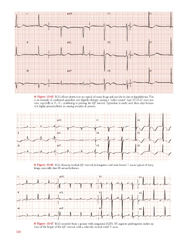
■ Figure 15-45 ECG effects shown here are typical of many drugs and can also be due to hypokalemia. This
is an example of combined quinidine and digitalis therapy causing a “roller coaster” type ST–T–U wave pat-
tern, especially in V 1 –V 3 , combining to prolong the QT interval. Quinidine is rarely used these days because
it is highly proarrhythmic in causing torsades de pointes.
V
aV
aV
V
V V V V V V V VR
V
a a a a a
V4
V4
V44
V V V V V V V V V V V
I I I I I aV R R R R R R R R R V V V V V V V V V V V V V V V 1 1 1 1 1 V4 4 4 4 4 4 4 4 4 4
aV
I I I I I I I I I aV L L L L L L L L L V V V V V V V V V V V V V V V 2 2 2 2 2 V5 5 5 5 5 5
aV
a a a a a
V5
V55
V V V V V V V V V V V
V5
V V V V V V V VL
V
V
V
a a a a a
aV
aV
V6
V V V V V V V V V V V
I I I I I I I I I I I I I aV F F F F F F F F F F V V V V V V V V V V V V V V V 3 3 3 3 3 V6 6 6 6 6 6 6 6 6 6
V66
V
V6
V V V V V V V VF
V
V
■ Figure 15-46 ECG showing marked QT interval prolongation and wide bizarre T waves typical of many
drugs, especially class III antiarrhythmics.
R
V1
V1
VR
V V V V V
VR
VR
I I I I a a a a a a aV R R R R R R R R V1 1 1 1 1 1 V V V V V V V V V V V V V V4 4 4 4 4
VRR
V V V V V V
V1
VL
I I I I I I I I a a a a a a aV L L L L L L L L L V V V V V V 2 2 2 2 2 2 2 2 2 2 V V V V V V V V V V V V V V5 5 5 5 5 5
V2
V2
V2
V2
VL
V V V V V
VL
VLL
VF
VFF
I I I I I I I I I a a a a a a aV F V3 3 3 3 3 3 3 3 3 3 3 V V V V V V V V V V V6 6 6 6 6
VF
V V V V
V3
V3
F F F F F F
V V V V V
■ Figure 15-47 ECG recorded from a patient with congenital LQTS. ST segment prolongation makes up
most of the length of the QT interval, with a relatively normal width T wave.
330