Page 349 - Cardiac Nursing
P. 349

LWBK340-c15_p300-332.qxd  6/29/09  10:30 PM  Page 325 Aptara Inc.


                                                                                C HAPTER 1 5 / Electrocardiography  325


                                             V V V V
                                             VR
                                            aV
                                            a a a a a a a a
                                            aVV
                                             VR
                                             V
                                             V
                                             V
                                             VR
                                             VR
                                              R
                                              R
                    I I I I I I I I I       aV R R R R R R R R        V1 1 1 1 1                V V V V V V V V V V4 4 4 4 4 4 4 4 4
                                                                      V V V V V V V V
                                                                      V1
                                                                      V1
                                            a a a a a a a a a a
                                            aV
                                             VL
                                            aVV
                                             VL
                                             V
                                             VL
                                             V
                                             VL
                                            aV
                                             V V V V
                                                                      V2
                                                                      V2
                                                                      V V V V V V V
                                                                      V2
                    I I I I II II II II I I I II I  aV L L L L L L L L L L  V2 2 2 2 2 2 2 2 2 2 2  V V V V V V V V V V5 5 5 5 5 5 5 5 5 5
                                             V
                                             V V V V
                                             VF
                                             V
                                             V
                                             VF
                                             VF
                                             VF
                                            aV
                                            a a a a a a a a
                                            aVV
                                                                      V3
                                                                      V V V V V V V
                                                                      V3
                                                                      V3
                     I I I I II II II II II II II II I I I I I I I  aV F F F F F F F F F F F F F F  V3 3 3 3 3 3 3 3 3 3 3  V V V V V V V V V V6 6 6 6 6 6 6 6 6 6
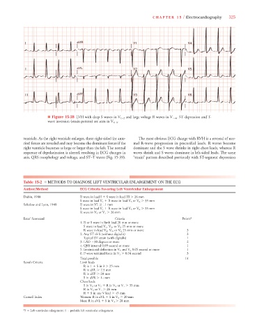
                              ■ Figure 15-38 LVH with deep S waves in V 1–2 and large voltage R waves in V 4–6 . ST depression and T-
                                                                                        V
                              wave inversion (strain pattern) are seen in V 4–6 .
                                                            V
                   ventricle. As the right ventricle enlarges, these right-sided (or ante-  The most obvious ECG change with RVH is a reversal of nor-
                   rior) forces are revealed and may become the dominant forces if the  mal R-wave progression in precordial leads. R waves become
                   right ventricle becomes as large or larger than the left. The normal  dominant and the S wave shrinks in right chest leads, whereas R
                   sequence of depolarization is altered, resulting in ECG changes in  waves shrink and S waves dominate in left-sided leads. The same
                   axis, QRS morphology and voltage, and ST–T waves (Fig. 15-39).  “strain” pattern described previously with ST-segment depression
                   Table 15-2  ■ METHODS TO DIAGNOSE LEFT VENTRICULAR ENLARGEMENT ON THE ECG
                   Author/Method              ECG Criteria Favoring Left Ventricular Enlargement
                   Dubin, 1988                R wave in lead I 
 S wave in lead III   26 mm
                                              S wave in lead V 1 
 R wave in lead V 5 or V 6   35 mm
                   Sokolow and Lyon, 1949     R wave in VL   11 mm
                                              S wave in lead V 1 
 R wave in lead V 5 or V 6   35 mm
                                              R wave in V 5 or V 6   26 mm
                   Estes’ Scorecard                           Criteria                        Points*
                                              1. R or S wave in limb lead 20 mm or more
                                                           V
                                               S wave in lead V 1 , V 2 , or V 3 25 mm or more
                                                         V
                                               R wave in lead V 4 , V 5 , or V 6 25 mm or more  3
                                              2. Any ST shift (without digitalis)               3
                                               Typical ST strain (with digitalis)               1
                                              3. LAD  30 degrees or more                        2
                                              4. QRS interval 0.09 second or more               1
                                              5. Intrinsicoid deflection in V 5 and V 6 0.05 second or more  1
                                              6. P-wave terminal force in V 1   0.04 second     3
                                              Total possible                                   14
                   Scott’s Criteria           Limb leads
                                               R in 1 
 S in 3   25 mm
                                               R in aVL   7.5 mm
                                               R in aVF   20 mm
                                               S in aVR   14 mm
                                              Chest leads
                                               S in V 1 or V 2 
 R in V 5 or V 6   35 mm
                                                      V
                                               R in V 5 or V 6   26 mm
                                               R 
 S in any V lead   45 mm
                   Cornell Index              Women: R in aVL 
 S in V 3   20 mm
                                              Men: R in aVL 
 S in V 3   28 mm
                   *5   Left ventricular enlargement; 4   probable left ventricular enlargement.
   344   345   346   347   348   349   350   351   352   353   354