Page 353 - Cardiac Nursing
P. 353

1
                                   0:3
                                   1
                                /09
                                /09
                                   0:3
                                        M
                                          Pa
                                        M
                                     0 P
                                     0 P
                          q
                          q
                          q
                        32.
                        32.
                           xd
                              /29
                              /29
                             6
                           xd
                             6
                                          Pa
                                                  t
                                                 p
                                                  t
                                                  ara
                                                  ara
                                                 p
                                              29
                                              29
                                                A
                                                 p
                                                A
                                            e 3
                                            e 3
                                           g
                                           g
                                           g
                                                      c.
                                                   a
                                                   a
                                                    In
                                                      c.
                                                    In
                    30
            K34
            K34
         LWB
         LWB
                    30
               0-c
                 15_
                 15_
                   p
                   p
               0-c
                      0-3
                      0-3
         LWBK340-c15_ pp300-332.qxd  6/29/09  10:30 PM  Page 329 Aptara Inc.
                                                                                C HAPTER 1 5 / Electrocardiography  329
                                                                       V1
                                                                       V1
                                                                       V1
                                                                       V V1
                                                                                                 V4
                                                                                                 V4
                                                                                                 V4
                                             aVR
                                                                                                 V V
                                               R
                                             aVR
                    I I I I I                aVR                       V 1                       V4 4 4
                                              V
                                             aVR
                                              V
                                             a aVR
                                             aVL
                                             aVL
                                             a aVL
                    III I II II              a aVL                     V2 2 2 2 2 2 2 2          V5 5 5 5
                                               L
                                              V
                                              V
                                                                       V2
                                                                                                 V V V5
                                                                                                 V5
                                                                       V V V2
                                                                       V3
                                             aVF
                                                                       V3 3
                                                                       V3
                                              V
                                                                       V V V3
                                              V
                                                                       V V V3
                                              V
                                                                                                 V V V6
                                                                                                 V6
                                             aVF FVFVFVVF
                                             aVF
                                                                                                 V V V6
                                                                                                 V6
                    III II I III II I III II I III III III  a aVF      V V V3 3 3 3 3 3 3 3 3 3 3 3 3 3 3 3  V6 6 6 6 6 6 6
                                                                                                 V V V6
                                             a aVF
                                             aVF
                                             aVF
                                             a aVF
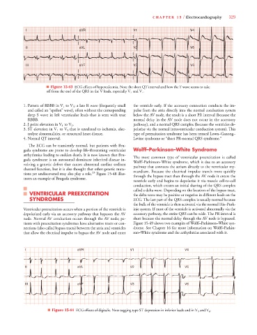
                              ■ Figure 15-43 ECG effects of hypercalcemia. Note the short QT interval and how the T wave seems to take
                              off from the end of the QRS in the V leads, especially V 3 and V 4 .
                                                                         V
                   1. Pattern of RBBB in V 1 to V 3 : a late R wave (frequently small  the ventricle early. If the accessory connection conducts the im-
                     and called an “epsilon” wave), often without the corresponding  pulse from the atria directly into the normal conduction system
                     deep S wave in left ventricular leads that is seen with true  below the AV node, the result is a short PR interval (because the
                     RBBB.                                             normal delay in the AV node does not occur in the accessory
                   2. J point elevation in V 1 to V 3 .                pathway), and a normal QRS complex (because the ventricles de-
                   3. ST elevation in V 1 to V 3 that is unrelated to ischemia, elec-  polarize via the normal intraventricular conduction system). This
                     trolyte abnormalities, or structural heart disease.  type of preexcitation syndrome has been termed Lown–Ganong–
                   4. Normal QT interval.                              Levine syndrome or “short PR-normal QRS syndrome.”
                     The ECG can be transiently normal, but patients with Bru-
                   gada syndrome are prone to develop life-threatening ventricular  Wolff–Parkinson–White Syndrome
                   arrhythmias leading to sudden death. It is now known that Bru-  The most common type of ventricular  preexcitation is called
                   gada syndrome is an autosomal dominant inherited disease in-  Wolff–Parkinson–White syndrome, which is due to an accessory
                   volving a genetic defect that causes abnormal cardiac sodium  pathway that connects the atrium directly to the ventricular my-
                   channel function, but it is also thought that other genetic muta-  ocardium. Because the electrical impulse travels more quickly
                   tions yet undiscovered may also play a role. 23  Figure 15-48 illus-  through the bypass tract than through the AV node it enters the
                   trates an example of Brugada syndrome.
                                                                       ventricle early and begins to depolarize it via muscle cell-to-cell
                                                                       conduction, which creates an initial slurring of the QRS complex
                                                                       called a delta wave. Depending on the location of the bypass tract,
                      VENTRICULAR PREEXCITATION                        the delta wave may be positive or negative in different leads on the
                      SYNDROMES                                        ECG. The last part of the QRS complex is usually normal because
                                                                       the bulk of the ventricle is then activated via the normal His–Purk-
                   Ventricular preexcitation occurs when a portion of the ventricle is  inje system. If most of the ventricle is activated abnormally via the
                   depolarized early via an accessory pathway that bypasses the AV  accessory pathway, the entire QRS can be wide. The PR interval is
                   node. Normal AV conduction occurs through the AV node; pa-  short because the normal delay through the AV node is bypassed.
                   tients with preexcitation syndromes have alternative tracts or con-  Figure 15-49 shows two examples of Wolff–Parkinson–White syn-
                   nections (also called bypass tracts) between the atria and ventricles  drome. See Chapter 16 for more information on Wolff–Parkin-
                   that allow the electrical impulse to bypass the AV node and enter  son–White syndrome and the arrhythmias associated with it.
                                                                     V1
                                                                     V1
                                                                     V1
                                                                                                4 4 4 4 4 4 4 4 4 4 4 4
                                                                     V V V V V V V V V V V V V V
                                             V
                                             VRR
                                             VR
                                             VR
                                             V
                                             V
                                             VR
                                             V V V V V V V V
                                            aV
                                            aV
                                              R
                    I I I I                 a a a a a a a a aV R R R R R R R R  V1 1 1 1 1 1 1  V V V V V V V V V V V V V V4
                                             VL
                                             V
                                             V
                                             VL
                                             VL
                                             VLL
                                             V V V V V V V V
                    II II II II             aV L L L L L L L L L     V2 2 2 2 2 2 2 2 2 2 2    V V V V V V V V V V V V V V5
                                            aV
                                            aV
                                             V
                                                                                                5 5 5 5 5 5 5 5 5 5 5 5
                                            a a a a a a a a
                                                                     V2
                                                                     V V V V V V V V V V V V V V
                                                                     V2
                                                                     V2
                                            aV
                                            a a a a a a a a
                                             V V V V V V V V
                                             V
                                                                                                6 6 6 6 6 6 6 6 6
                                             V
                                            aV
                     I I I I II II II II    aV F F F F F F F F F F F F F F  V3 3 3 3 3 3 3 3 3  V V V V V V V V V V V V V V6
                                             V
                                             V
                                             V
                                             V
                                             V
                                             V
                                             V
                                             V
                                             V
                                                                     V V3
                                                                     V V V V V V V V V V V V V V
                                                                     V3
                                             V
                                             V
                                             V
                                             V
                                             V
                                             V
                                             V
                                             V
                              ■ Figure 15-44 ECG effects of digitalis. Note sagging type ST depression in inferior leads and in V 5 and V 6 .
   348   349   350   351   352   353   354   355   356   357   358