Page 407 - Cardiac Nursing
P. 407

Pa
                                          Pa
                                        M
                                        M
                                           g
                                            e 3
                                            e 3
                                           g
                                           g
                                /09
                                   1
                                                      c.
                                /09
                                   1
                                     6 A
                                     6 A
                                   2:1
                                   2:1
                                                  ara
                                                   a
                                                  t
                                                  ara
                                                   a
                                                      c.
                                                      c.
                                                     In
                                                     In
                                                A
                                                A
                                              83
                                              83
                                                A
                                                 p
                                                  t
                                                 p
                                                 p
                          q
                          q
                        87.
                      3-3
                        87.
                              6
                             6
                           xd
                          q
                           xd
                      3-3
            K34
               0-c
         LWBK340-c16_
         LWB K34 0-c 16_ pp333-387.qxd  6/30/09  12:16 AM  Page 383 Aptara Inc.
         LWB
                    33
                    33
                   p
                 16_
                   p
                              /30
                              /30
                             6
                                                             C HAPTER 1 6 / Arrhythmias and Conduction Disturbances  383
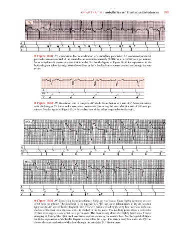
                           V 1 1
                              ■ Figure 16-37 AV dissociation due to acceleration of a subsidiary pacemaker. An accelerated junctional
                              pacemaker assumes control of the ventricles and conducts aberrantly (RBBB) at a rate of 88 beats per minute.
                              Sinus arrhythmia is present at a rate that is in the 70s. See the legend of Figure 16-36 for explanation of the
                              ladder diagram below the strip. Vertical wavy lines in the V level indicate aberrant conduction through the ven-
                              tricles.
                                    A
                                    AV
                                    V
                              ■ Figure 16-38 AV dissociation due to complete AV block. Sinus rhythm at a rate of 65 beats per minute
                              with third-degree AV block and a ventricular pacemaker controlling the ventricles at a rate of 38 beats per
                              minute. See the legend of Figure 16-36 for explanation of the ladder diagram below the strip.


                              ■ Figure 16-39 AV dissociation due to interference. Strips are continuous. Sinus rhythm is present at a rate
                              of 68 beats per minute. The third beat in the top strip is a PJC that causes refractoriness in the AV junction
                              (gray area in AV level of ladder diagram). The refractory period created by the early beat interferes with con-
                              duction of the next sinus impulse, which is blocked in the AV node. The resulting pause allows a ventricular
                              rhythm to emerge at a rate of 65 beats per minute. The bottom strip shows the slightly faster sinus P waves
                              emerging in front of the QRS until ventricular capture occurs in the seventh beat. See the legend of Figure
                              16-36 for explanation of the ladder diagram drawn below the strips. The vertical wavy line under the PJC in-
                              dicates aberrant conduction of that beat through the ventricles. F   fusion beats.
   402   403   404   405   406   407   408   409   410   411   412