
Page 405 - Cardiac Nursing
P. 405
Pa
Pa
M
M
Pa
g
e 3
g
g
6 A
/09
1
/09
/09
1
2:1
6 A
1
2:1
ara
a
t
ara
a
c.
c.
In
In
t
81
81
e 3
81
A
p
p
A
p
3-3
3-3
33
q
87.
87.
33
LWBK340-c16_
LWB
LWB K34 0-c 16_ p p pp333-387.qxd 6/30/09 12:16 AM Page 381 Aptara Inc.
16_
0-c
K34
6
xd
6
/30
q
/30
xd
q
C HAPTER 1 6 / Arrhythmias and Conduction Disturbances 381
V 1 1
A
V V 1
B
A
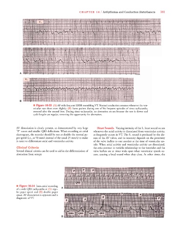
■ Figure 16-33F 6 33 (A) AF with frequent LBBB resembling VT. Normal conduction resumes whenever the ven-
tricular rate slows even slightly. (B) Same patient during one of his frequent episodes of sinus tachycardia,
restored after the second beat. During sinus tachycardia, no aberration occurs because the rate is slower and
cycle lengths are regular, removing the opportunity for aberration.
AV dissociation is clearly present, as demonstrated by very large Heart Sounds. Varying intensity of the S 1 heart sound occurs
“P” waves and smaller QRS deflections. When recording an atrial whenever the atrial activity is dissociated from ventricular activity,
electrogram, the recorder should be run at double the normal pa- as frequently occurs in VT. The S 1 sound is produced by the clo-
per speed (i.e., at 50 mm/s instead of the usual 25 mm/s) to make sure of the AV valves, and its intensity depends on the proximity
it easier to differentiate atrial and ventricular activity. of the valve leaflets to one another at the time of ventricular sys-
tole. When atrial activity and ventricular activity are dissociated,
Clinical Criteria the atria contract in variable relationship to the ventricles and the
Several clinical criteria can be used to aid in the differentiation of valve leaflets are at times wide open when ventricular systole oc-
aberration from ectopy. curs, causing a loud sound when they close. At other times, the
■ Figure 16-34 Intra-atrial recording
A
of a wide QRS tachycardia at (A) regu-
lar paper speed and (B) double paper
speed. AV dissociation is apparent and is
diagnostic of VT.
B B