
Page 468 - Cardiac Nursing
P. 468
1:2
9 P
1:2
1
1
9 P
Pa
Pa
Pa
M
M
1
xd
6
xd
q
q
6
/09
/09
/09
/29
/29
g
a
a
ara
t
ara
a
c.
c.
In
a
In
t
e 4
44
e 4
g
g
44
p
p
p
A
A
LWB K34 0-c 20_ p p pp439-459.qxd 6/29/09 11:29 PM Page 444 Aptara Inc.
LWB
43
43
LWBK340-c20_
20_
20_
K34
0-c
59.
9-4
9-4
q
59.
59.
444 P A R T III / Assessment of Heart Disease
A B C
D E F
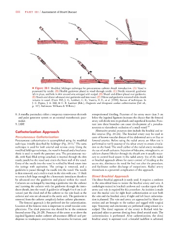
■ Figure 20-1 Modified Seldinger technique for percutaneous catheter sheath introduction. (A) Vessel is
punctured by needle. (B) Flexible guidewire placed in vessel through needle. (C) Needle removed, guidewire
left in place, and hole in skin around wire enlarged with scalpel. (D) Sheath and dilator placed over guidewire.
(E) Sheath and dilator advanced over guidewire and into vessel. (F) Dilator and guidewire removed while sheath
remains in vessel. (From Hill, J. A., Lambert, C. R., Vuestra, R. E., et al. [1998]. Review of techniques. In
J. C. Pepine, J. A. Hill, & C. R. Lambert [Eds.], Diagnostic and therapeutic cardiac catheterization [3rd ed.,
p. 107]. Baltimore: Williams & Wilkins.)
8. A standby pacemaker, either a temporary transvenous electrode retroperitoneal bleeding. Puncture of the artery more than 3 cm
and pulse generator system or an external transthoracic pace- below the inguinal ligament increases the chance that the femoral
maker. artery will divide into its profunda and superficial branches. Punc-
9. IABP. ture into these branches can cause development of a pseudoa-
neurysm or thrombotic occlusion of a small vessel. 20
Catheterization Approach Alternative arterial puncture sites include the brachial and ra-
dial arteries (Fig. 20-2A). The brachial artery may be used in
Percutaneous Catheterization cases of known vascular disease of the abdominal aorta or iliac or
Percutaneous catheterization is accomplished using the modified femoral arteries. Before using the radial artery, an Allen test is
5
technique initially described by Seldinger (Fig. 20-1). The same performed to verify patency of the ulnar artery to ensure circula-
technique is used for both arterial and venous entry. Using the tion to the hand. The small caliber of the radial artery mandates
modified Seldinger technique, the vessel is located and a local anes- the use of small catheters. Injection of lidocaine, nitroglycerin, or
thetic is used to numb the puncture area. The percutaneous nee- calcium channel blocker through the sheath arm is usually neces-
dle, with fluid-filled syringe attached, is inserted through the skin sary to control local spasm in the radial artery. Use of the radial
nearly parallel to the vessel and enters the front wall of the vessel. or brachial approach allows for easier control of bleeding at the
Entry of the needle into the vessel is verified by blood return into access site, eliminates the need for bed rest after the procedure,
the syringe with aspiration. The syringe is removed, and a and facilitates earlier discharge of outpatients. Radial artery
guidewire is passed through the needle into the vessel. The needle thrombosis is a potential complication of this approach.
is then removed, and a nick is made in the skin with a no. 11 blade
to create a hole large enough for a hemostatic introducer sheath to Direct Brachial Approach
be advanced over the guidewire and placed within the vessel. The direct brachial approach is rarely used. It requires a cutdown
Catheters are exchanged by inserting a guidewire into the catheter in the antecubital fossa to isolate the brachial artery and vein. A
and inserting the catheter with the guidewire through the intro- cardiologist trained in brachial cutdown and vascular repair of the
ducer sheath, into the vessel. A guidewire of length 4 to 6 cm is ad- artery and vein is required for this procedure. An incision is made
vanced past the distal end of the catheter so the wire leads as the over the medial vein for right heart catheterization or over both
catheter and wire are advanced to the aortic arch. The guidewire is the vein and the brachial artery if right and left heart catheteriza-
removed from the catheter completely before catheter placement. tion is planned. The vein and artery are approached by blunt dis-
The femoral approach is the preferred site for catheterization. section and are brought to the surface and tagged with surgical
Location of the femoral stick is important to avoid vascular com- tape. Venotomy and arteriotomy are performed using scissors or a
plications. The ideal puncture site should be in the common scalpel. The distal segment of the artery is flushed with he-
femoral artery (Fig. 20-2B). Puncture of the artery at or above the parinized saline to prevent clotting from distal arterial stasis. The
inguinal ligament makes catheter advancement difficult and pre- catheterization is performed. After catheterization, the distal
disposes to inadequate compression, hematoma formation, and brachial artery is aspirated until a forceful backflow is achieved,