
Page 539 - Cardiac Nursing
P. 539
0 A
1:0
M
0 A
1
009
1:0
1
M
51
51
g
g
Pa
M
g
Pa
q
q
36.
q
3
3
xd
xd
36.
6/2
0/0
009
6/2
1-5
1-5
0/0
3
e 5
ara
LWB K34 0-c 22_ p p pp511-536.qxd 30/06/2009 11:00 AM Page 515 Aptara
ara
ara
LWB
0-c
22_
LWBK340-c22_
K34
t
15
A
e 5
15
A
p
t
p
p
C HAPTER 22 / Acute Coronary Syndromes 515
A
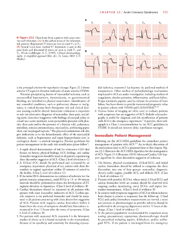
V 1 V 2 V 3 V 4 V 5
■ Figure 22-2 Chest leads from a patient with acute ante-
rior wall infarction. (A) In the earliest phase of the infarction,
V
tall, positive (hyperacute) T waves are seen in leads V 2 to V 5 .
(B) Several hours later, marked ST depression is seen in the
same leads and abnormal Q waves are seen in leads V 1 and B
V 2 . (From Goldberger, A. L. [1999]. Clinical electrocardiog-
V V 1 V 2 V 3 V 4 V 5
raphy: A simplified approach [6th ed.]. St. Louis, MO: C.V.
Mosby.)
is the principal criterion for reperfusion therapy. Figure 22-2 shows dial ischemia, troponin-I has become the preferred method of
anterior ST-segment elevation indicative of acute anterior STEMI. measurement. Other markers of pathophysiologic mechanisms
Potential precipitating factors of myocardial ischemia, such as implicated in ACS are under investigation, including markers of
uncontrolled hypertension, thyrotoxicosis, or gastrointestinal coagulation, platelet activation, inflammation, and heart failure.
bleeding, are identified on physical examination. Identification of B-type natriuretic peptide, used to indicate the presence of heart
any comorbid conditions, such as pulmonary disease or malig- failure, has been shown to provide incremental prognostic value
nancy, is critical because both therapeutic risk and clinical deci- in patient cohorts with STEMI and UA/NSTEMI. 8–10
sion making could be altered. Immediate evaluation is important 6. Various forms of imaging are often used to evaluate patients
to rule out alternative diagnoses of noncardiac chest pain, includ- with symptoms that are suggestive of ACS. Bedside echocardio-
ing aortic dissection (suggestive with findings of unequal pulses or graphy is useful for diagnosis and risk stratification of patients
2
a loud new aortic murmur), acute pericarditis (presents with pleu- with ACS in the emergency department. A portable chest radi-
ritic chest pain and/or the presence of a friction rub), pulmonary ograph is a Class I recommendation by the ACC guidelines in
embolism, tension pneumothorax, myocarditis, perforating peptic STEMI. It should not however delay reperfusion strategies.
2
ulcer, and esophageal rupture. The physical examination will also
give indications as to the hemodynamic effect of the myocardial Immediate Patient Management
ischemia, such as hypotension and organ hypoperfusion seen in
cardiogenic shock—a medical emergency. Practice guidelines for Following are the ACC/AHA guidelines for immediate patient
4
patient management in the early risk stratification phase follow : management of patients with ACS. 2,4 An in-depth discussion of
the medications used in ACS is presented later in this chapter. Fig-
1. A rapid clinical determination of risk for obstructive CAD that
ure 22-3 illustrates the ACC/AHA algorithm for the management
focuses on history, physical findings, ECG findings, and cardiac
of ACS. Figure 22-4 illustrates AHA’s Advanced Cardiac Life Sup-
biomarker integration should be made in all patients experiencing
port algorithm for chest discomfort suggestive of ischemia.
chest discomfort suggestive of ACS. (Class I, level of evidence: C)
2. A 12-lead ECG should be performed and evaluated by an 1. The history, physical examination, 12-lead ECG, and initial
emergency department physician of any patient with chest dis- cardiac biomarkers should be utilized to categorize the chest
comfort or anginal equivalent within 10 minutes of arrival to discomfort into one of four groups: noncardiac diagnosis,
the facility. (Class I, level of evidence: C) chronic stable angina, possible ACS, and definite ACS. (Class
3.If the initial ECG demonstrates no evidence of ischemia but the I, level of evidence: C)
patient remains symptomatic, serial ECGs at 15- to 30-minute 2. Patients with possible ACS but whose initial 12-lead ECG and
intervals should be performed to detect the development of ST- cardiac biomarker levels are normal should be observed with
segment elevation or depression. (Class I, level of evidence: B) ongoing cardiac monitoring, serial ECGs, and repeat bio-
4. Cardiac biomarkers should be measured in all patients who marker measurement. (Class I, level of evidence: B)
present with chest discomfort indicative of ACS. Cardiac-spe- 3. In patients with symptoms suggestive of ACS in whom ischemic
cific troponin is the preferred biomarker and should be meas- heart disease is present or suspected, but the follow-up 12-lead
ured in all patients presenting with chest discomfort suggestive ECG and cardiac biomarkers measurements are normal, a stress
of ACS. Patients with negative cardiac biomarkers within 6 test (exercise or pharmacologic) to provoke ischemia should be
hours from the onset of symptoms should have biomarkers re- performed in the emergency department within 72 hours of dis-
measured in 8 to 12 hours after the onset of symptoms. (Class charge. (Class I, level of evidence: C)
I, level of evidence: B) 4. In the patient population recommended for outpatient stress
5. For patients with suspected ACS, troponin-I is the laboratory testing, precautionary appropriate pharmacotherapy should
marker of choice as it is found exclusively in the myocardium. be prescribed including aspirin, -blockers, and/or sublin-
Because of its specificity and sensitivity for detecting myocar- gual NTG. If the patient is discharged from the emergency