Page 538 - Cardiac Nursing
P. 538

M
                                          M
                                        0 A
                                        0 A
                                            Pa
                                              g
                                          M
                                            Pa
                                  009
                                  009
                                6/2
                                6/2
                                      1:0
                                      1:0
                                     1
                                     1
                                                   p
                                                    t
                                                   p
                                                   p
                                                     ara
                                                     ara
                                                    t
                                                     ara
                                               e 5
                                               e 5
                                              g
                                              g
                                                  A
                                                  A
                                                14
                                                14
                          q
                           xd
                          q
                        36.
                          q
                           xd
                              0/0
                              0/0
                             3
                              3
                             3
            K34
               0-c
         LWB
         LWB K34 0-c 22_ p p pp511-536.qxd  30/06/2009  11:00 AM  Page 514 Aptara
         LWBK340-c22_
                 22_
                      1-5
                        36.
                      1-5
                    51
                    51
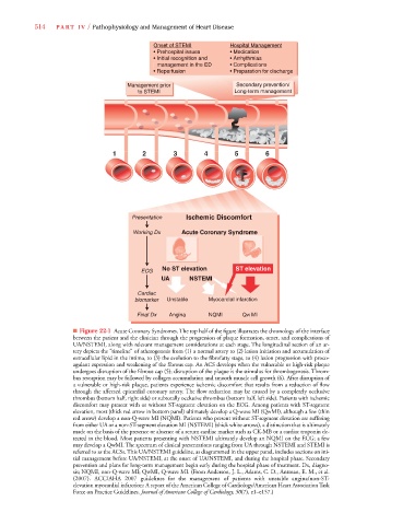
                  514    PA R T  I V / Pathophysiology and Management of Heart Disease
                                                      Onset of STEMI         Hospital Management
                                                                                p
                                                                                       g
                                                      • Prehospital issues   • Medication
                                                      • Initial recognition and  • Arrhythmias
                                                         management in the ED  • Complications
                                                      • Reperfusion          • Preparation for discharge
                                              Management prior                 Secondary prevention/
                                                 to STEMI                     Long-term management
                                          1        2        3        4        5         6
                                               Presentation     Ischemic Discomfort
                                                Working Dx    Acute Coronary Syndrome
                                                         No ST elevation       ST elevation
                                                  ECG
                                                        UA       NSTEMI
                                                 Cardiac
                                                biomarker  Unstable   Myocardial infarction
                                                 Final Dx  Angina     NQMI       QW MI
                              ■ Figure 22-1 Acute Coronary Syndromes. The top half of the figure illustrates the chronology of the interface
                              between the patient and the clinician through the progression of plaque formation, onset, and complications of
                              UA/NSTEMI, along with relevant management considerations at each stage. The longitudinal section of an ar-
                              tery depicts the “timeline” of atherogenesis from (1) a normal artery to (2) lesion initiation and accumulation of
                              extracellular lipid in the intima, to (3) the evolution to the fibrofatty stage, to (4) lesion progression with proco-
                              agulant expression and weakening of the fibrous cap. An ACS develops when the vulnerable or high-risk plaque
                              undergoes disruption of the fibrous cap (5); disruption of the plaque is the stimulus for thrombogenesis. Throm-
                              bus resorption may be followed by collagen accumulation and smooth muscle cell growth (6). After disruption of
                              a vulnerable or high-risk plaque, patients experience ischemic discomfort that results from a reduction of flow
                              through the affected epicardial coronary artery. The flow reduction may be caused by a completely occlusive
                              thrombus (bottom half, right side) or subtotally occlusive thrombus (bottom half, left side). Patients with ischemic
                              discomfort may present with or without ST-segment elevation on the ECG. Among patients with ST-segment
                              elevation, most (thick red arrow in bottom panel) ultimately develop a Q-wave MI (QwMI), although a few (thin
                              red arrow) develop a non-Q-wave MI (NQMI). Patients who present without ST-segment elevation are suffering
                              from either UA or a non-ST-segment elevation MI (NSTEMI) (thick white arrows), a distinction that is ultimately
                              made on the basis of the presence or absence of a serum cardiac marker such as CK-MB or a cardiac troponin de-
                              tected in the blood. Most patients presenting with NSTEMI ultimately develop an NQMI on the ECG; a few
                              may develop a QwMI. The spectrum of clinical presentations ranging from UA through NSTEMI and STEMI is
                              referred to as the ACSs. This UA/NSTEMI guideline, as diagrammed in the upper panel, includes sections on ini-
                              tial management before UA/NSTEMI, at the onset of UA/NSTEMI, and during the hospital phase. Secondary
                              prevention and plans for long-term management begin early during the hospital phase of treatment. Dx, diagno-
                              sis; NQMI, non-Q-wave MI; QwMI, Q-wave MI. (From Anderson, J. L., Adams, C. D., Antman, E. M., et al.
                              (2007). ACC/AHA 2007 guidelines for the management of patients with unstable angina/non-ST-
                              elevation myocardial infarction: A report of the American College of Cardiology/American Heart Association Task
                              Force on Practice Guidelines. Journal of American College of Cardiology, 50(7), e1–e157.)
   533   534   535   536   537   538   539   540   541   542   543