
Page 582 - Cardiac Nursing
P. 582
LWBK340-c24_p555-594.qxd 30/06/2009 01:43 PM Page 558 Aptara
558 PA R T IV / Pathophysiology and Management of Heart Disease
cease replicating and dividing early in life. Myocardial insult and
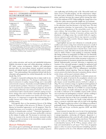
Table 24-2 ■ ESTABLISHED AND HYPOTHESIZED RISK injury occurs as decades progress, and the compensatory adapta-
FACTORS FOR HEART FAILURE tion often leads to dysfunction. Preventing myocyte hypertrophy,
injury, and death becomes the primary goal in altering the trajec-
Major Risk Toxic Minor
Factors Precipitants Risk Factors tory of the syndrome of HF. Understanding the changes that occur
within the cardiac myocyte provides insight into the syndrome.
Asymptomatic LV Chemotherapy Smoking Increased pressure or volume reactivates growth factors present
dysfunction (anthracyclines, Dyslipidemia in the embryonic heart but dormant in the adult heart. This fetal
Increased LV mass cyclophosphamide, Sleep-disordered
Age, male gender 5-FU, trastuzumab) breathing gene expression stimulates the hypertrophy of the myocytes and
Hypertension, LVH Cocaine Chronic renal disease the synthesis and degradation of the extracellular matrix. There is
Myocardial infarction NSAIDs Anemia some evidence that extracellular matrix degradation may elicit
Diabetes Doxazosin Sedentary lifestyle side-to-side slippage or disarray of myocytes, perhaps caused by
Valvular heart disease Alcohol Low socioeconomic dissolution of collagen struts that normally hold cells together,
Obesity status
Psychological stress whereas reparative and reactive fibrosis may represent a secondary
event resulting in a stiffer ventricle. Myocyte slippage may also be
5-FU, 5-florouracil; LV, left ventricle; LVH, left ventricle hypertrophy; NSAIDs, non- caused by myocyte loss. 13
steroidal anti-inflammatory agents. Hypertrophied myocytes have alterations in contractile protein
Adapted from Schocken, D. D., Benjamin, E. J., Fonarow, G. C., et al. [2008]. Preven-
tion of heart failure. A scientific statement from the American Heart Association synthesis and calcium cycling. Force within the myocyte is a result of
Councils on Epidemiology and Prevention, Clinical Cardiology, Cardiovascular Nurs- the interaction of myosin and actin. Myocyte hypertrophy alters the
ing, and High Blood Pressure Research; Quality of Care and Outcomes Research In- synthesis of myocin proteins from -myosin heavy chain toward
terdisciplinary Working Group; and Functional Genomics and Translational Biology
Interdisciplinary Working Group. Circulation, Table 1. -myosin heavy chain. This shift in the myosin subunit alters the ki-
netics by which it binds to actin resulting in contraction. Initially,
this change produces an energetically favorable state, but chronically
this state is unsustainable and failure occurs. Altered expression or
alignment of contractile proteins within sarcomeres is important, as
increasing mutations in sarcomeric proteins have been linked to in-
and cytokine activation, and vascular and endothelial dysfunction. herited cardiomyopathic processes. Alteration in expression and
Multiple alterations in organ and cellular physiology contribute to function of the contractile proteins is signaled by mechanical wall
HF under various circumstances. Adaptive and maladaptive stress, angiotensin II (AT), norepinephrine (NE), endothelin (ET),
processes affect the myocardium, kidneys, peripheral vasculature, tumor necrosis factor-alpha (TNF- ), inflammatory interleukins,
smooth and skeletal muscle, and multiple reflex control mecha- and intracellular calcium signaling. There are changes in the sar-
11
nisms. Our understanding of the mechanism behind both the de- comeric proteins that lead to decreased contraction velocity, reduced
velopment and progression has evolved dramatically over the last stroke volume, and increased ventricular volume. 7,11
decade (Fig. 24-3). Ventricular contraction and relaxation is a dynamic process con-
Changes in myocardial architecture due to injury result in my- trolled by the uptake of calcium by the sarcoplasmic reticulum and
ocardial contractile dysfunction. The changes in architecture come the efflux of calcium within the myocyte. 11–13 Myocyte hypertro-
about by alterations in cardiac myocyte biology, and in myocardial phy impacts several intracellular proteins involved in calcium cy-
and ventricular structures. Ventricular contractile dysfunction leads cling. The most consistent change is a significant downregulation of
to altered systemic perfusion. Alterations in systemic perfusion re- expression and activity of the sarcoplasmic reticulum calcium ATP-
sult in neuroendocrine activation resulting in progressive alterations ase (SERCA) pump. While the role of SERCA in calcium handling
in myocardial architecture and contractile function. In short, my- within the myoctye is complex and remains the topic of study, the
ocardial injury leads to altered systemic perfusion causing neuroen- net result is a reduction in the availability of peak systolic calcium,
dorine changes that result in further ventricular dysfunction. The and an elevation and prolongation in diastolic calcium, resulting in
remainder of this section examines more closely each of the steps in reduced systolic contraction and delayed diastolic relaxation.
this process. It is important to recognize that understanding the in- The paracrine function of the heart is markedly altered in HF.
terplay between each of these steps is a dynamic and rapidly ex- Paracrine action is the release of a locally acting endocrine sub-
panding venture. stance, a system in which the target cells are close to the signaling
Histologically there are four prominent features of the failing cells. The signal transduction system in the failing myocardium is
heart: (1) hypertrophy of the myocytes, (2) fibrosis, (3) myocyte profoundly altered. The failing myocardium is induced to secrete
disarray (or unordered appearance), and (4) apoptosis. These his- both atrial and b-type natriuretic peptides (ANP and BNP,
tological processes occur secondary to myocardial ischemia, in- respectively).
farction, or hemodynamic overload. 12 Ventricular hypertrophy is Actual myocyte loss may also occur by one of two mechanisms:
defined by an increase in ventricular mass attributable to increase apoptosis or necrosis. Apoptosis is a programmed cell death that
in the volume of cardiac cells. The increase in ventricular mass is is energy-dependent, producing cell dropout. Apoptosis is a
due to an increase in the size of the myocytes, increased number highly regulated process that causes the cell to shrink, yielding cell
of fibroblasts, and an increase in the extracellular matrix proteins fragments that are surrounded by plasma membrane. This process
(collagen and fibronectin). does not invoke an inflammatory or fibrotic response. Apoptosis
is stimulated by hypoxia, AT II, TNF- , myocyte calcium over-
Myocyte Pathophysiology load, and mitochondrial or cell injury. 14 Two forms of apoptosis
The primary goal in preventing and ultimately managing HF is appear to affect the course of postinfarction remodeling: ischemic-
protection and preservation of the myocyte. Cardiac myocytes driven apoptosis at the site of the infarction and load-dependent