
Page 585 - Cardiac Nursing
P. 585
0/0
6/2
0/0
3
3
6/2
0
1:4
0
009
009
3
5-5
94.
5-5
55
55
94.
xd
xd
q
q
q
A
p
A
61
61
p
ara
ara
t
p
t
e 5
M
M
3 P
1:4
3 P
Pa
g
e 5
g
Pa
g
LWB
LWBK340-c24_
LWB K34 0-c 24_ p p pp555-594.qxd 30/06/2009 01:43 PM Page 561 Aptara
24_
0-c
K34
C HAPTER 24 / Heart Failure and Cardiogenic Shock 561
positive-feedback loop can be generated in which impaired pump
performance increases impedance to LV ejection, further impair-
ing pump performance.
Changes in the composition of the myocardium occur in re-
sponse to injury or overload and result in structural remodeling,
divided into both cellular and noncellular changes. Several of the
cellular changes have be discussed in the prior section and include
changes within the cardiomyocytes (hypertrophy, apoptosis, and
necrosis) but several alterations in other cell types, such as fibrob-
lasts, vascular smooth muscle cells, monocytes and macrophages,
also contribute to ventricular remodeling. Noncellular compo-
nents of the myocardium likewise contribute to remodeling.
There is an increase in interstitial deposition of collagen fibers and
an increase in perivascular deposition of collagen, leading to
thickening of the walls of the small intramyocardial arteries and
arterioles.
Throughout the body there is a fine balance of synthesis and
degradation. Fibroblasts within the myocardium produce collagen.
Excesses in ventricular collagen are thought to be caused by both an
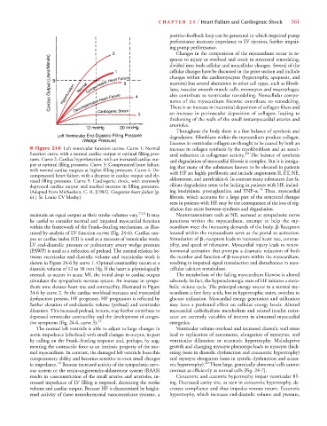
■ Figure 24-6 Left ventricular function curves. Curve 1: Normal increase in collagen synthesis by the myofibroblasts and an associ-
function curve, with a normal cardiac output at optimal filling pres- ated reduction in collagenase activity. 19 The balance of synthesis
sures. Curve 2: Cardiac hyperfunction, with an increased cardiac out- and degradation of myocardial fibrosis is complex. But it is intrigu-
put at optimal filling pressures. Curve 3: Compensated heart failure, ing that many of the substances known to be elevated in patients
with normal cardiac outputs at higher filling pressures. Curve 4: De-
compensated heart failure, with a decrease in cardiac output and ele- with HF are highly profibrotic and include angiotensin II, ET, NE,
vated filling pressures. Curve 5: Cardiogenic shock, with extremely aldosterone, and interleukin-6. In contrast many substances that fa-
depressed cardiac output and marked increase in filling pressures. cilitate degradation seem to be lacking in patients with HF, includ-
(Adapted from Michaelson, C. R. [1983]. Congestive heart failure [p. ing bradykinin, prostaglandins, and TNF- . 11 Thus, myocardial
61]. St. Louis: CV Mosby.) fibrosis, which accounts for a large part of the structural changes
seen in patients with HF, may be the consequence of the loss of reg-
ulation that exists between synthesis and degradation.
maintain an equal output as their stroke volumes vary. 7,11 It may Neurotransmitters such as NE, secreted at sympathetic nerve
be useful to consider normal and impaired myocardial function junctions within the myocardium, attempt to help the my-
within the framework of the Frank–Starling mechanism, as illus- ocardium meet the increasing demands of the body. -Receptors
trated by analysis of LV function curves (Fig. 24-6). Cardiac out- located within the myocardium serve as the portal to activation.
put or cardiac index (CI) is used as a measure of ventricular work; Stimulation of 1 -receptors leads to increased heart rate, contrac-
LV end-diastolic pressure or pulmonary artery wedge pressure tility, and speed of relaxation. Myocardial injury leads to neuro-
(PAWP) is used as a reflection of preload. The normal relation be- hormonal activation that prompts a dramatic reduction of both
tween ventricular end-diastolic volume and ventricular work is the number and function of -receptors within the myocardium,
shown in Figure 24-6 by curve 1. Optimal contractility occurs at a resulting in impaired signal transduction and disturbances in myo-
diastolic volume of 12 to 18 mm Hg. If the heart is physiologically cellular calcium metabolism.
stressed, as occurs in acute MI, the initial drop in cardiac output The metabolism of the failing myocardium likewise is altered
stimulates the sympathetic nervous system. An increase in sympa- adversely. In fact, the hyperadrenergic state of HF initiates a meta-
thetic tone elevates heart rate and contractility, illustrated in Figure bolic vicious cycle. The principal energy source in a normal my-
24-6 by curve 2. As the cardiac workload increases and myocardial ocardium is free fatty acids, but in hypertrophic states, switches to
dysfunction persists, HF progresses. HF progression is reflected by glucose utilization. Myocardial energy generation and utilization
further elevation of end-diastolic volume (preload) and ventricular may have a profound effect on cellular energy levels. Altered
dilatation. This increased preload, in turn, may further contribute to myocardial carbohydrate metabolism and related insulin resist-
depressed ventricular contractility and the development of conges- ance are currently variables of interest in abnormal myocardial
tive symptoms (Fig. 24-6, curve 3). 17 energetics.
The normal left ventricle is able to adjust to large changes in Ventricular volume overload and increased diastolic wall stress
aortic impedance (afterload) with small changes in output, in part lead to replication of sarcomeres, elongation of myocytes, and
by calling on the Frank–Starling response and, perhaps, by aug- ventricular dilatation or eccentric hypertrophy. Maladaptive
menting the contractile force as an intrinsic property of the nor- growth and changing myocyte phenotype leads to myocyte thick-
mal myocardium. In contrast, the damaged left ventricle loses this ening (seen in diastolic dysfunction and concentric hypertrophy)
compensatory ability and becomes sensitive to even small changes and myocyte elongation (seen in systolic dysfunction and eccen-
18
20
in impedance. Because increased activity of the sympathetic nerv- tric hypertrophy). These large, genetically abnormal cells cannot
ous system or the renin–angiotensin–aldosterone system (RAAS) contract as efficiently as normal cells (Fig. 24-7).
results in vasoconstriction of the small arteries and arterioles, in- Concentric and eccentric hypertrophy impair ventricular fill-
creased impedance of LV filling is imposed, decreasing the stroke ing. Decreased cavity size, as seen in concentric hypertrophy, de-
volume and cardiac output. Because HF is characterized by height- creases compliance and thus impedes venous return. Eccentric
ened activity of these neurohormonal vasoconstrictor systems, a hypertrophy, which increases end-diastolic volume and pressure,