
Page 586 - Cardiac Nursing
P. 586
6/2
009
6/2
0/0
0/0
1:4
1:4
0
009
0
3
q
q
94.
5-5
94.
3
3
xd
q
xd
3 P
p
p
A
62
A
ara
ara
t
p
t
62
Pa
Pa
M
3 P
M
e 5
e 5
g
g
g
0-c
24_
K34
LWB
LWBK340-c24_
LWB K34 0-c 24_ p p pp555-594.qxd 30/06/2009 01:43 PM Page 562 Aptara
5-5
55
55
562 PA R T I V / Pathophysiology and Management of Heart Disease
Physiologic Athlete's heart
hypertrophy
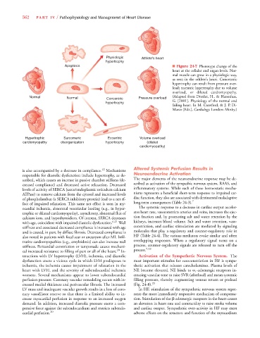
Apoptosis ■ Figure 24-7 Phenotype change of the
heart at the cellular and organ levels. Nor-
mal muscle can grow in a physiologic way,
as seen in the athlete’s heart. Concentric
hypertrophy can result from pressure over-
load; eccentric hypertrophy due to volume
overload, or dilated cardiomyopathy.
Normal Normal Concentric Pressure overload (Adapted from Drexler, H., & Hasenfuss,
hypertrophy G. [2001]. Physiology of the normal and
failing heart. In M. Crawford, & J. P. Di-
Marco [Eds.], Cardiology. London: Mosby.)
Hypertrophic Sarcomeric Eccentric Volume overload
cardiomyopathy disorganization hypertrophy (dilated
cardiomyopathy)
is also accompanied by a decrease in compliance. 12 Mechanisms Altered Systemic Perfusion Results in
responsible for diastolic dysfunction include hypertrophy, as de- Neuroendocrine Activation
scribed, which causes an increase in passive chamber stiffness (de- The major elements of the neuroendocrine response may be de-
creased compliance) and decreased active relaxation. Decreased scribed as activation of the sympathic nervous system, RAAS, and
levels of activity of SERCA (sarco/endoplasmic reticulum calcium inflammatory systems. While each of these homeostatic mecha-
ATPase) to remove calcium from the cytosol and increased levels nisms represents a beneficial short-term response to impaired car-
of phospholamban (a SERCA inhibitory protein) lead to a net ef- diac function, they also are associated with detrimental maladaptive
7
fect of impaired relaxation. This same net effect is seen in my- long-term consequences (Table 24-3).
ocardial ischemia, abnormal ventricular loading (e.g., in hyper- The systemic response to a decrease in cardiac output acceler-
trophic or dilated cardiomyopathy), asynchrony, abnormal flux of ates heart rate, vasoconstricts arteries and veins, increases the ejec-
calcium ions, and hypothyroidism. Of interest, SERCA decreases tion fraction and, by promoting salt and water retention by the
with age, coincident with impaired diastolic dysfunction. 6,21 Wall kidneys, increases blood volume. Salt and water retention, vaso-
stiffness and associated decreased compliance is increased with age constriction, and cardiac stimulation are mediated by signaling
and is caused, in part, by diffuse fibrosis. Decreased compliance is molecules that play a regulatory and counter-regulatory role in
also noted in patients with focal scar or aneurysm after MI. Infil- HF (Table 24-4). The various mediators evoke similar and often
trative cardiomyopathies (e.g., amyloidosis) can also increase wall overlapping responses. When a regulatory signal turns on a
stiffness. Pericardial constriction or tamponade causes mechani- process, counter-regulatory signals are released to turn off the
7,22
cal increased resistance to filling of part or all of the heart. 20 In- process.
teractions with LV hypertrophy (LVH), ischemia, and diastolic Activation of the Sympathetic Nervous System. The
dysfunction create a vicious cycle in which LVH predisposes to most important stimulus for vasoconstriction in HF is sympa-
ischemia, the ischemia causes impairment of relaxation in the thetic activation that releases catecholamines. Plasma levels of
heart with LVH, and the severity of subendocardial ischemia NE become elevated. NE binds to 1 -adrenergic receptors in-
worsens. Several mechanisms appear to lower subendocardial creasing vascular tone to raise SVR (afterload) and mean systemic
perfusion pressure. Coronary vascular remodeling occurs with in- filling pressure, thereby augmenting venous return or preload
creased medial thickness and perivascular fibrosis. The increased (Fig. 24-8). 23
LV mass and inadequate vascular growth results in a loss of coro- In HF, stimulation of the sympathetic nervous system repre-
nary vasodilator reserve so that there is a limited ability to in- sents the most immediately responsive mechanism of compensa-
crease myocardial perfusion in response to an increased oxygen tion. Stimulation of the -adrenergic receptors in the heart causes
demand. In addition, increased diastolic pressure exerts a com- an elevation in heart rate and contractility to raise stroke volume
pressive force against the subendocardium and restricts subendo- and cardiac output. Sympathetic over-activity in HF may exert
cardial perfusion. 20 adverse effects on the structure and function of the myocardium