
Page 592 - Cardiac Nursing
P. 592
LWBK340-c24_p555-594.qxd 30/06/2009 01:43 PM Page 568 Aptara
568 PA R T IV / Pathophysiology and Management of Heart Disease
is of particular benefit for specifically assessing ventricular mass,
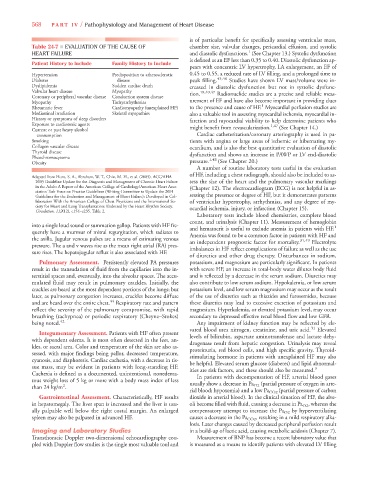
Table 24-7 ■ EVALUATION OF THE CAUSE OF chamber size, valvular changes, pericardial effusion, and systolic
1
HEART FAILURE and diastolic dysfunctions. (See Chapter 13.) Systolic dysfunction
is defined as an EF less than 0.35 to 0.40. Diastolic dysfunction ap-
Patient History to Include Family History to Include
pears with concentric LV hypertrophy, LA enlargement, an EF of
Hypertension Predisposition to atherosclerotic 0.45 to 0.55, a reduced rate of LV filling, and a prolonged time to
Diabetes disease peak filling. 45,46 Studies have shown LV mass/volume were in-
Dyslipidemia Sudden cardiac death creased in diastolic dysfunction but not in systolic dysfunc-
Valvular heart disease Myopathy 18,40,47
Coronary or peripheral vascular disease Conduction system disease tion. Radionuclide studies are a precise and reliable meas-
Myopathy Tachyarrhythmias urement of EF and have also become important in providing clues
1
Rheumatic fever Cardiomyopathy (unexplained HF) to the presence and cause of HF. Myocardial perfusion studies are
Mediastinal irradiation Skeletal myopathies also a valuable tool in assessing myocardial ischemia, myocardial in-
History or symptoms of sleep disorders farction and myocardial viability to help determine patients who
Exposure to cardiotoxic agents 1,47
Current or past heavy alcohol might benefit from revascularization. (See Chapter 14.)
consumption Cardiac catheterization/coronary arteriography is used in pa-
Smoking tients with angina or large areas of ischemic or hibernating my-
Collagen vascular disease ocardium, and is also the best quantitative evaluation of diastolic
Thyroid disease
Pheochromocytoma dysfunction and shows an increase in PAWP or LV end-diastolic
1,48
Obesity pressure. (See Chapter 20.)
A number of routine laboratory tests useful in the evaluation
of HF, including a chest radiograph, should also be included to as-
Adapted from Hunt, S. A., Abraham, W. T., Chin, M. H., et al. (2005). ACC/AHA
2005 Guideline Update for the Diagnosis and Management of Chronic Heart Failure sess the size of the heart and the pulmonary vascular markings
in the Adult: A Report of the American College of Cardiology/American Heart Asso- (Chapter 12). The electrocardiogram (ECG) is not helpful in as-
ciation Task Force on Practice Guidelines (Writing Committee to Update the 2001
Guidelines for the Evaluation and Management of Heart Failure): Developed in Col- sessing the presence or degree of HF, but it demonstrates patterns
laboration With the American College of Chest Physicians and the International So- of ventricular hypertrophy, arrhythmias, and any degree of my-
ciety for Heart and Lung Transplantation: Endorsed by the Heart Rhythm Society. ocardial ischemia, injury, or infarction (Chapter 15).
Circulation, 112(12), e154–e235. Table 2.
Laboratory tests include blood chemistries, complete blood
count, and urinalysis (Chapter 11). Measurement of hemoglobin
into a single loud sound or summation gallop. Patients with HF fre- 1
and hematocrit is useful to exclude anemia in patients with HF.
quently have a murmur of mitral regurgitation, which radiates to
Anemia was found to be a common factor in patients with HF and
the axilla. Jugular venous pulses are a means of estimating venous 31,49
an independent prognostic factor for mortality. Electrolyte
pressure. The a and v waves rise as the mean right atrial (RA) pres-
imbalances in HF reflect complications of failure as well as the use
sure rises. The hepatojugular reflux is also associated with HF.
of diuretics and other drug therapy. Disturbances in sodium,
Pulmonary Assessment. Persistently elevated PA pressures potassium, and magnesium are particularly significant. In patients
result in the transudation of fluid from the capillaries into the in- with severe HF, an increase in total-body water dilutes body fluid
terstitial spaces and, eventually, into the alveolar spaces. The accu- and is reflected by a decrease in the serum sodium. Diuretics may
mulated fluid may result in pulmonary crackles. Initially, the also contribute to low serum sodium. Hypokalemia, or low serum
crackles are heard at the most dependent portions of the lungs; but potassium level, and low serum magnesium may occur as the result
later, as pulmonary congestion increases, crackles become diffuse of the use of diuretics such as thiazides and furosemides, because
44
and are heard over the entire chest. Respiratory rate and pattern these diuretics may lead to excessive excretion of potassium and
reflect the severity of the pulmonary compromise, with rapid magnesium. Hyperkalemia, or elevated potassium level, may occur
breathing (tachypnea) or periodic respiratory (Cheyne–Stokes) secondary to depressed effective renal blood flow and low GFR.
being noted. 42 Any impairment of kidney function may be reflected by ele-
vated blood urea nitrogen, creatinine, and uric acid. 31 Elevated
Integumentary Assessment. Patients with HF often present
levels of bilirubin, aspartate aminotransferase and lactate dehy-
with dependent edema. It is most often detected in the feet, an-
drogenase result from hepatic congestion. Urinalysis may reveal
kles, or sacral area. Color and temperature of the skin are also as-
proteinuria, red blood cells, and high specific gravity. Thyroid-
sessed, with major findings being pallor, decreased temperature,
stimulating hormone in patients with unexplained HF may also
cyanosis, and diaphoresis. Cardiac cachexia, with a decrease in tis-
be helpful. Elevated serum glucose (diabetes) and lipid abnormal-
sue mass, may be evident in patients with long-standing HF. 9
ities are risk factors, and these should also be measured.
Cachexia is defined as a documented, unintentional, nonedema-
In patients with decompensation of HF, arterial blood gases
tous weight loss of 5 kg or more with a body mass index of less
2
than 24 kg/m . usually show a decrease in Pa O2 (partial pressure of oxygen in arte-
rial blood; hypoxemia) and a low Pa CO2 (partial pressure of carbon
Gastrointestinal Assessment. Characteristically, HF results dioxide in arterial blood). In the clinical situation of HF, the alve-
in hepatomegaly. The liver span is increased and the liver is usu- oli become filled with fluid, causing a decrease in Pa O2 , whereas the
ally palpable well below the right costal margin. An enlarged compensatory attempt to increase the Pa O2 by hyperventilating
spleen may also be palpated in advanced HF. causes a decrease in the Pa CO2 , resulting in a mild respiratory alka-
losis. Later changes caused by decreased peripheral perfusion result
Imaging and Laboratory Studies in a build-up of lactic acid, causing metabolic acidosis (Chapter 7).
Transthoracic Doppler two-dimensional echocardiography cou- Measurement of BNP has become a recent laboratory value that
pled with Doppler flow studies is the single most valuable tool and is measured as a means to identify patients with elevated LV filling