
Page 603 - Cardiac Nursing
P. 603
LWBK340-c24_p555-594.qxd 30/06/2009 01:43 PM Page 579 Aptara
C HAP TE R 24 / Heart Failure and Cardiogenic Shock 579
occurs in less than 1% of acute MIs but carries a high mortality Extracardiac Obstructive Shock
rate. 195
Non-MI-related acute valvular problems involve the mitral and Pericardial Tamponade
aortic valve. Acute mitral regurgitation can be caused by sponta- The accumulation of fluid within the pericardial sac increases
neous chordal rupture, infective endocarditis, inflammatory disor- pressure, causing extracardiac obstruction to filling that results in a
ders (e.g., rheumatic fever), or trauma. Acute aortic insufficiency decrease in ventricular preload and cardiac output. What deter-
may be caused by infective endocarditis with leaflet destruction mines whether pericardial effusion will cause shock is how rapidly
(most common), acute aortic dissection, or traumatic injury. the fluid accumulates. Patients at risk for shock caused by tampon-
Shock may be caused by aortic stenosis with increasing metabolic ade are those with malignancy (especially lung and breast cancer,
demands or with concomitant LV failure. Mitral stenosis rarely lymphoma, leukemia, or melanoma), infection, aortic dissection,
causes shock without rapid atrial fibrillation. 195 Prosthetic valve or severe pericarditis.
dysfunction, especially left-sided, most often causes shock because
of valvular insufficiency. Acute prosthetic valvular insufficiency Pulmonary Embolism
occurs because of dehiscence of the sewing ring, infective endo- When embolic material, such as thrombus, fat, tumor, or air, ob-
carditis, or catastrophic mechanical failure. structs 30% or more or the pulmonary vasculature, the RV can-
Infiltrative disease, such as amyloidosis, sarcoidosis, and he- not provide adequate pressure to compensate for the increased re-
mochromatosis, are examples of infiltrative diseases in their later sistance to blood flow. RV failure ensues, with increased RV
stages that may be associated with shock. Shock caused by trauma end-diastolic and RA pressures, and finally a decrease in cardiac
is usually seen secondary to myocardial or aortic rupture, or output and shock.
caused by acute volume loss secondary to hemorrhage.
Acute decompensation of chronic HF represents a somewhat Compensatory Mechanisms
different pathophysiologic state, because these patients have a
marked reduction in LV systolic function at baseline as com- The following equations illustrate the physiologic relation of the
pared to those patients with acute HF without prior LV dys- hemodynamic variables. Here CO, cardiac output; SV, stroke
function. 190 Patients with chronic HF are likely to be using volume; HR, heart rate; MAP, mean arterial pressure; and SVR,
combination therapy, usually an ACE-I, diuretic, -blocker, systemic vascular resistance compose the equations:
and/or digoxin. There is already activation of the neurohor-
monal compensatory mechanisms, including increased sympa- CO SV HR
thetic stimulation of the heart, activation of the RAAS, increased MAP CO SVR
vasoconstriction, fluid retention by the kidneys, increased ven- In the pathophysiologic state of cardiogenic shock, the de-
tricular preload, and LV hypertrophy and remodeling. When a crease in MAP is brought about by an alteration in one of the vari-
precipitating event occurs, there is further derangement of ables. The reduction in cardiac output results from a decrease in
these compensatory mechanisms. Factors leading to acute de- stroke volume:
compensation in chronic HF may include the following: acute
myocardial ischemia, poorly treated or untreated hypertension, TCO TSV HR
new-onset atrial fibrillation, concurrent infections (e.g., pneu- The deduction in MAP results from the decrease in cardiac
monia, influenza), medication noncompliance, excess dietary output:
sodium, cardiac depressant drugs, NSAIDs, and endocrine ab-
normalities (e.g., poorly controlled diabetes, hyperthy- TMAP TCO SVR
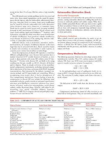
roidism). 190 Table 24-13 compares clinical and pathophysiologic Compensatory mechanisms consist of reflex reactions to an
features of acute and chronic HF. initial fall in blood pressure. They are activated immediately and
Table 24-13 ■ COMPARISON OF ACUTE AND CHRONIC HEART FAILURE
Decompensated Chronic Stable Chronic
Clinical Feature Acute Heart Failure Heart Failure Heart Failure
Symptom severity (shortness of breath Marked and sudden Moderate to severe None to Mild or moderate
and fatigue)
Pulmonary edema Common Frequent Rare
Peripheral edema Rare Frequent Occasional
Weight gain None to mild Very frequent Occassional
Total body volume No change to mild increase Marked increase Mild increase
Cardiomegaly Uncommon Common Common
LV systolic function Hypo-, normo- or hypercontractile Normal to reduced Normal to reduced
LV wall stress Marked increase Marked increase Elevated
Activation of sympathetic nervous system Marked increase Marked increase Mild to marked increase
Activation of RAAS Marked increase Marked increase Mild to marked increase
Myocardial ischemia* Common Occasional Rare
Hypertensive crisis Common Occasional Rare
*For example, acute coronary syndrome, acute mitral regurgitation, aortic stenosis, or ventricular septal defect.
LV, left ventricle; RAAS, renin–angiotensin–aldosterone system.