
Page 684 - Cardiac Nursing
P. 684
7
/1/
09
qxd
p65
5-7
04.
660
Ap
tar
g
49
AM
P
0-c
28_
K34
LWB K34 0-c 28_ p65 5-7 04. qxd 7 /1/ 09 9: 49 AM P a a g e e 660 Ap tar a a
LWB
LWBK340-c28_p655-704.qxd 7/1/09 9:9:49 AM Page 660 Aptara
660 PA R T I V / Pathophysiology and Management Disease
DISPLAY 28-3 Dual Chamber Pacemaker Terminology (continued)
cardia that results when there is retrograde conduction from the ventricle to the atria, sensing of the retrograde P wave
by the atrial channel, and pacing in the ventricle in response to the sensed P wave. This results in a reentry tachycardia
in which the pacemaker serves as the antegrade limb of the circuit and the intrinsic conduction system serves as the ret-
rograde limb. Also known as endless loop tachycardia or pacemaker reentry tachycardia.
Psuedopseudofusion beat: An electrocardiographic phenomenon in which an atrial pacing spike is superimposed on a
native QRS complex. The atrial pacing spike cannot contribute to ventricular depolarization, but the presence of the spike
can distort the native QRS complex on the ECG.
Postventricular atrial refractory period (PVARP): Part of the total atrial refractory period that begins with a sensed or
paced ventricular event. PVARP is a programmable parameter and is intended to prevent the atrial channel from sensing
far-field ventricular signals, such as T waves or local myocardial potentials. PVARP can also be programmed to prevent
the atrial channel from sensing retrograde P waves, thus preventing PMT.
Rate drop response: Pacing at a rate faster than the programmed pacing rate when the patient’s intrinsic heart rate drops
suddenly, as in vasovagal syncope or hypersensitive carotid sinus syndrome. Pacing is initiated at a rate up to 110 bpm if
bradycardia suddenly occurs.
Rate response: Ability of the pacemaker to increase its pacing rate in response to physical activity or increased metabolic
demand. Rate responsive pacemakers have some type of sensor that detects physical activity or a physiological parame-
ter that indicates the need for increased heart rate. Currently, the sensors most commonly used are vibration or motion
sensors and minute ventilation sensors. Other sensors being evaluated include blood temperature, blood oxygen content,
QT interval, and stroke volume. Also known as rate modulation or rate adaptation.
Rate smoothing: A programmable function that prevents excessive cycle-to-cycle changes in pacing rate. Atrial tracking and
rate response can occur but no sudden acceleration or deceleration in pacing rate can occur.
Safety pacing: The delivery of a ventricular output at a short AV interval whenever a signal is sensed early in the AV delay.
The purpose of safety pacing is to prevent crosstalk inhibition of ventricular output. Also known as nonphysiological AV
delay or ventricular safety standby.
Sleep rate: The pacemaker is programmed to gradually decrease the base pacing rate to a lower limit (e.g., a sleep rate of
50 bpm) at bedtime and gradually increase the base pacing rate when the patient awakens.
Total atrial refractory period (TARP): Timing cycle that determines the total length of time that the atrial channel is unre-
sponsive to signals (in effect “has its eyes closed”). TARP is composed of two separately programmable timing cycles
during which the atrial channel is refractory: the AV interval and PVARP.
Ventricular refractory period: The amount of time following a ventricular sensed or paced event during which the ventricu-
lar channel cannot respond to signals (in effect “has its eyes closed”). The purpose is to prevent the ventricular channel
from seeing large repolarization signals (T waves) or other local myocardial signals.
formed in emergency and elective situations, and it is usually per- the apex of the right ventricle for ventricular pacing, the right
formed in a monitored unit such as critical care or telemetry unit. atrium for atrial pacing, or both chambers for dual-chamber pac-
Transcutaneous pacing can also be performed by paramedics or ing (Fig. 28-2). The transvenous pacing lead is attached to an ex-
other trained personnel in emergency response vehicles or in the ternal pulse generator that is kept either on the patient or at the
field. bedside. The procedure is usually performed under fluoroscopy in
a cardiac catheterization laboratory but it can be done at the bed-
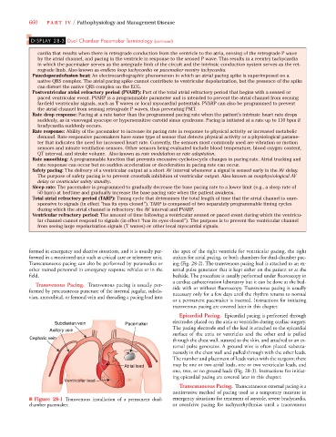
Transvenous Pacing. Transvenous pacing is usually per-
side with or without fluoroscopy. Transvenous pacing is usually
formed by percutaneous puncture of the internal jugular, subcla-
necessary only for a few days until the rhythm returns to normal
vian, antecubital, or femoral vein and threading a pacing lead into
or a permanent pacemaker is inserted. Instructions for initiating
transvenous pacing are covered later in this chapter.
Epicardial Pacing. Epicardial pacing is performed through
Subclavian vein Pacemaker electrodes placed on the atria or ventricles during cardiac surgery.
The pacing electrode end of the lead is attached to the epicardial
Axillary vein
surface of the atria or ventricles and the other end is pulled
Cephalic vein n n through the chest wall, sutured to the skin, and attached to an ex-
ternal pulse generator. A ground wire is often placed subcuta-
neously in the chest wall and pulled through with the other leads.
The number and placement of leads varies with the surgeon; there
Atrial lea ad may be one or two atrial leads, one or two ventricular leads, and
ad
a a a
ad
d d d d d d d
ad
ad
one, two, or no ground leads (Fig. 28-3). Instructions for initiat-
ing epicardial pacing are covered later in this chapter.
cular
ntricular lead
ul
cula
entricular lead
Ve
V Ventricentricular leaentricular lead d
Ventricular
Ventricular le
lead
lead
lead
l
lead
ar
ul
ar
ar
Transcutaneous Pacing. Transcutaneous external pacing is a
noninvasive method of pacing used as a temporary measure in
■ Figure 28-1 Transvenous installation of a permanent dual- emergency situations for treatment of asystole, severe bradycardia,
chamber pacemaker. or overdrive pacing for tachyarrhythmias until a transvenous