
Page 765 - Cardiac Nursing
P. 765
CHAPTER 31 / Adult Congenital Heart Disease 741
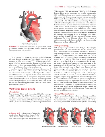
VSD, muscular VSD, and subarterial VSD (Fig. 31-6). Perimem-
branous defects are the commonest type of defect accounting for
80% of all VSDs and occur in the membranous part of the ventric-
ular septum and the surrounding muscular septum. A muscular
VSD occurs in the muscular portion of the septum and accounts for
approximately 15%. Subarterial VSDs occur in only 5% of cases
and are located directly below the atrioventricular valves. VSDs are
further categorized as either restrictive or unrestrictive. Restrictive
VSDs are small defects resulting in a high-pressure gradient be-
tween right and left ventricle, while nonrestrictive VSDs are large
defects in which the pressure between right and left ventricle is
equalized. Unrestricted defects are typically repaired in childhood,
but restrictive VSDs account for 7% of congenital heart defects
found in adults. Isolated VSD occur with equal frequency in men
and women. The 25 year follow-up indicates that the majority of
patients managed medically or surgically, who do not develop
Eisenmenger syndrom will fare well. 24,25
Pathophysiology
n Figure 31-5 Ventricular septal defect. (Reprinted from Everett,
A. PedHeart Resource. 2009. Scientific Software Solutions. www. Defects may be single or multiple, with the degree of shunting de-
heartpassport.com., with permission.) pendent on the size of the defect rather than the anatomic loca-
tion. Defects vary in size from a millimeter or two in diameter to
large openings with little or no septal wall, which behave physio-
logically like a single ventricle. Small isolated defects (,7 mm in
Today, transcatheter closure of ASD is the established method diameter), and moderate-sized defects (7 mm to 1.25 cm) are con-
of closure for patients with secundum ASD with success rates of sidered to be restrictive. They have minimal hemodynamic
greater than 95% being reported. 22,23 While occurring infre- changes and produce little or no symptomatology. But if moder-
quently, the long-term concerns for these devices include incom- ate in size, it may be significant enough to produce some cardiac
plete closure of the shunt, acute embolization of the devices, and enlargement. If the defect is large (1.5 to 3 cm), systemic pressure
the potential for thromboembolic complications. in the right ventricle is equal or slightly lower than the left ven-
Adults with simple ASD repaired in infancy and no residual ef- tricle creating a left-to-right shunt and increased pulmonary
fects may be followed at the community level. Patients with resid- blood flow (unrestricted VSD). This increase in blood flow is re-
ual lesions or patients in whom the ASD is diagnosed in adulthood turned to the left heart, creating volume overload of both right
should be evaluated at a regional ACHD center. Indications for and left ventricles. In addition, increased pulmonary blood flow
specialty care include pre- or postoperative arrhythmias, valvular may produce pulmonary hypertension. Because of the open com-
and/or ventricular dysfunction, and elevated pulmonary pressures. munication between the two ventricles, systolic blood pressure in
Endocarditis prophylaxis is only indicated within the first 6 the pulmonary artery rises, equaling that in the aorta. If the pul-
months after percutaneous closure with a device or surgical closure monary artery pressure continues to increase and pulmonary vas-
using prosthetic material. 20 cular resistance approaches or exceeds systemic pressure, shunt
reversal (right to left) occurs, rendering the patient cyanotic and
Ventricular Septal Defects no longer a candidate for surgical correction. This syndrome is
referred to as Eisenmenger reaction.
Description
Interventricular defects result in shunting of blood between right Clinical Manifestations
and left ventricles (Fig. 31-5). Ventricular septal defects (VSDs) Clinical features depend on the volume of pulmonary blood flow,
are the most common heart defects, representing about 35% of all which in turn depends on the size of the defect and the pulmonary
congenital cardiac anomalies. 4,14 There are three main categories vascular resistance. A harsh holosystolic murmur and palpable
of VSD, each classified according to its location: perimembranous thrill along the lower left sternal border may be the only findings
n Figure 31-6 Different types of ventricular
septal defects. (Reprinted from Everett, A. Ped-
Heart Resource. 2009. Scientific Software Solu-
tions. www.heartpassport.com., with permission.)