
Page 767 - Cardiac Nursing
P. 767
CHAPTER 31 / Adult Congenital Heart Disease 743
the base of the heart and is transmitted to the suprasternal notch
and over both carotid arteries. The typical murmur of valvular
aortic stenosis is a harsh, loud systolic murmur that begins after
the first heart sound, rising to a peak (crescendo) and declining
(decrescendo) before the second heart sound. The murmur radi-
ates to the suprasternal notch and carotid arteries. A systolic ejec-
tion sound, which may be heard at the cardiac apex, implies a mo-
bile valve and is found in mild to moderate stenosis. As
calcification impairs valve mobility, the ejection sound decreases
or vanishes completely.
Management
Asymptomatic patients with mild aortic stenosis and gradients
,25 mm Hg may be treated medically. With higher gradients
.50 mm Hg, balloon valvuloplasty may be successfully per-
formed resulting in a 60% to 70% reduction in systolic gradient
across the aortic valve. Balloon valvuloplasty is not recommended
if aortic insufficiency is present, in patients with subvalvar steno-
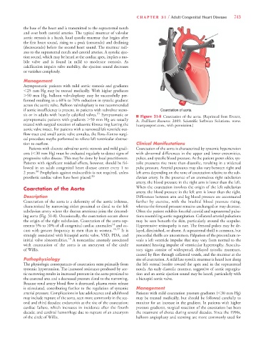
sis or in adults with heavily calcified valves. 21 Symptomatic or n Figure 31-8 Coarctation of the aorta. (Reprinted from Everett,
asymptomatic patients with gradients .50 mm Hg are usually A. PedHeart Resource. 2009. Scientific Software Solutions. www.
treated with surgical resection of subaortic fibrous ring leaving the heartpassport.com., with permission.)
aortic valve intact. For patients with a narrowed left ventricle out-
flow tract and small aortic valve annulus, the Ross–Konno surgi-
cal procedure maybe performed to relieve left ventricular obstruc-
tion to outflow. Clinical Manifestations
Patients with discrete subvalvar aortic stenosis and mild gradi- Coarctation of the aorta is characterized by systemic hypertension
ents (,30 mm Hg) must be evaluated regularly to detect signs of with abnormal differences in the upper and lower extremities,
progressive valve disease. This may be done by local practitioners. pulses, and systolic blood pressure. As the patient grows older, sys-
Patients with significant residual effects, however, should be fol- tolic pressures rise more than diastolic, resulting in a widened
lowed in an adult congenital heart disease center every 1 to pulse pressure. Arterial pressures may also vary between right and
2 years. 26 Prophylaxis against endocarditis is not required, unless left arms depending on the zone of coarctation relative to the sub-
prosthetic cardiac valves have been placed. 20 clavian artery. In the presence of an anomalous right subclavian
artery, the blood pressure in the right arm is lower than the left.
Coarctation of the Aorta When the coarctation involves the origin of the left subclavian
artery, the blood pressure in the left arm is lower than the right.
Description Differences between arm and leg blood pressure are accentuated
Coarctation of the aorta is a deformity of the aortic isthmus, further by exercise, with the brachial blood pressure rising,
characterized by narrowing either proximal or distal to the left whereas the femoral pressure remains unchanged or may decrease.
subclavian artery where the ductus arteriosus joins the descend- Often the patient exhibits forceful carotid and suprasternal pulsa-
ing aorta (Fig. 31-8). Occasionally, the coarctation occurs above tions resembling aortic regurgitation. Collateral arterial pulsations
the origin of the right subclavian. Coarctation of the aorta rep- may be seen beneath the skin, particularly around the scapulae.
resents 5% to 10% of all congenital cardiac anomalies 14 and oc- Hypertensive retinopathy is rare. The femoral pulses may be de-
curs with greater frequency in men than in women. 13,27 It is layed, diminished, or absent. A suprasternal thrill is common, but
strongly associated with bicuspid aortic valve, VSD, PDA, and precordial thrills are uncommon. Palpation of the precordium re-
initial valve abnormalities. 13 A noncardiac anomaly associated veals a left ventricle impulse that may vary from normal to the
with coarctation of the aorta is an aneurysm of the circle sustained heaving impulse of ventricular hypertrophy. Ausculta-
of Willis. tory signs consist of widespread, delayed systolic murmurs,
caused by flow through collateral vessels, and the murmur at the
Pathophysiology site of coarctation. A mild late systolic murmur is heard best along
The physiologic consequences of coarctation stem primarily from the left sternal border toward the apex and in the suprasternal
systemic hypertension. The increased resistance produced by aor- notch. An early diastolic murmur, suggestive of aortic regurgita-
tic narrowing results in increased pressure in the aorta proximal to tion and an aortic ejection sound may be heard, particularly with
the coarcted area and a decreased pressure distal to the narrowing. a bicuspid aortic valve.
Because renal artery blood flow is decreased, plasma renin release
is stimulated, contributing further to the regulation of systemic Management
arterial pressure. Complications in late adolescence and adulthood Patients with mild coarctation pressure gradients (,30 mm Hg)
may include rupture of the aorta, seen more commonly in the sec- may be treated medically, but should be followed carefully to
ond and third decades; endarteritis at the site of the coarctation; monitor for an increase in the gradient. In patients with higher
cardiac failure, which increases in incidence after the fourth pressure gradients, surgical resection of the coarctation has been
decade; and cerebral hemorrhage due to rupture of an aneurysm the treatment of choice during several decades. Since the 1990s,
of the circle of Willis. balloon angioplasty and stenting are more commonly used for