
Page 766 - Cardiac Nursing
P. 766
742 P AR T IV / Pathophysiology and Management of Heart Disease
of a small or moderate defect. A normal splitting second sound in-
dicates the pulmonary arterial pressure is below systemic pressure.
In large shunts, the murmur is lower in intensity, a mid-diastolic
“flow murmur” and third heart sound are heard at the apex, and a
right ventricular impulse is palpable.
Management
Patients with small VSD (,7 mm in diameter) have minimal he-
modynamic changes and produce little or no symptoms. There-
fore, they do not require surgical intervention. In the absence of
high or fixed pulmonary vascular obstructive disease, surgical cor-
rection is indicated in patients with moderate to large left-to-right
shunt. VSD closure will be considered if the Q p /Q s is higher than
2:1. According to the 2007 guidelines, endocarditis prophylaxis is
no longer indicated, except for patients with prosthetic material
(e.g. patch for VSD closure) with residua or within the first 6
months after an operation in which prosthetic material is
placed. 20
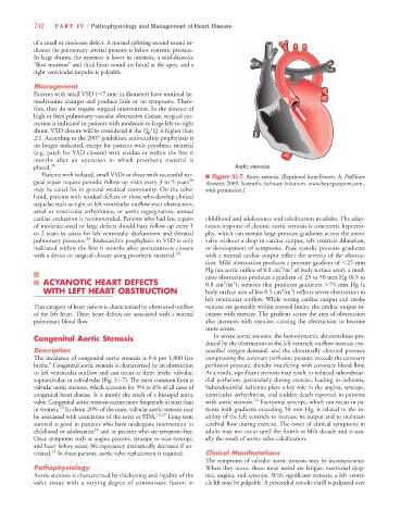
Patients with isolated, small VSDs or those with successful sur- n Figure 31-7 Aortic stenosis. (Reprinted from Everett, A. PedHeart
gical repair require periodic follow-up visits every 3 to 5 years 26 Resource. 2009. Scientific Software Solutions. www.heartpassport.com.,
may be cared for in general medical community. On the other with permission.)
hand, patients with residual defects or those who develop clinical
sequelae such as right or left ventricular outflow tract obstruction,
atrial or ventricular arrhythmias, or aortic regurgitation, annual
cardiac evaluation is recommended. Patients who had late repairs childhood and adolescence and calcification in adults. The adap-
of moderate-sized or large defects should have follow-up every 1 tation response of chronic aortic stenosis is concentric hypertro-
to 2 years to assess for left ventricular dysfunction and elevated phy, which can sustain large pressure gradients across the aortic
pulmonary pressures. 26 Endocarditis prophylaxis in VSD is only valve without a drop in cardiac output, left ventricle dilatation,
indicated within the first 6 months after percutaneous closure or development of symptoms. Peak systolic pressure gradients
with a device or surgical closure using prosthetic material. 20 with a normal cardiac output reflect the severity of the obstruc-
tion. Mild obstruction produces a pressure gradient of ,25 mm
2
2
Hg (an aortic orifice of 0.8 cm /m of body surface area); a mod-
erate obstruction produces a gradient of 25 to 50 mm Hg (0.5 to
ACYANOTIC HEART DEFECTS 0.8 cm /m ); stenosis that produces gradients .75 mm Hg (a
2
2
WITH LEFT HEART OBSTRUCTION body surface area of less 0.5 cm /m ) reflects severe obstruction to
2
2
left ventricular outflow. While resting cardiac output and stroke
This category of heart defects is characterized by obstructed outflow volume are generally within normal limits, the cardiac output in-
of the left heart. These heart defects are associated with a normal creases with exercise. The gradient across the area of obstruction
pulmonary blood flow. also increases with exercise, causing the obstruction to become
more severe.
Congenital Aortic Stenosis In severe aortic stenosis, the hemodynamic abnormalities pro-
duced by the obstruction to the left ventricle outflow increase my-
Description ocardial oxygen demand, and the abnormally elevated pressure
The incidence of congenital aortic stenosis is 0.4 per 1,000 live compressing the coronary perfusion pressure exceeds the coronary
4
births. Congenital aortic stenosis is characterized by an obstruction perfusion pressure, thereby interfering with coronary blood flow.
to left ventricular outflow and can occur at three levels: valvular, As a result, significant stenosis may result in reduced subendocar-
supravalvular, or subvalvular (Fig. 31-7). The most common form is dial perfusion, particularly during exercise, leading to ischemia.
valvular aortic stenosis, which accounts for 3% to 6% of all cases of Subendocardial ischemia plays a key role in the angina, syncope,
congenital heart disease. It is mostly the result of a bicuspid aortic ventricular arrhythmias, and sudden death reported in patients
valve. Congenital aortic stenosis occurs more frequently in men than with aortic stenosis. 14 Exertional syncope, which can occur in pa-
13
in women. In about 20% of the cases, valvular aortic stenosis may tients with gradients exceeding 50 mm Hg, is related to the in-
be associated with coarctation of the aorta or PDA. 13,27 Long-term ability of the left ventricle to increase its output and to maintain
survival is good in patients who have undergone intervention in cerebral flow during exercise. The onset of clinical symptoms in
childhood or adolescence 28 and in patients who are symptom-free. adults may not occur until the fourth or fifth decade and is usu-
Once symptoms such as angina pectoris, syncope or near-syncope, ally the result of aortic valve calcification.
and heart failure occur, life expectancy dramatically decreases if un-
13
treated. In these patients, aortic valve replacement is required. Clinical Manifestations
The symptoms of valvular aortic stenosis may be inconspicuous.
Pathophysiology When they occur, those most noted are fatigue, exertional dysp-
Aortic stenosis is characterized by thickening and rigidity of the nea, angina, and syncope. With significant stenosis, a left ventri-
valve tissue with a varying degree of commissure fusion in cle lift may be palpable. A precordial systolic thrill is palpated over