
Page 206 - ACCCN's Critical Care Nursing
P. 206
Cardiovascular Assessment and Monitoring 183
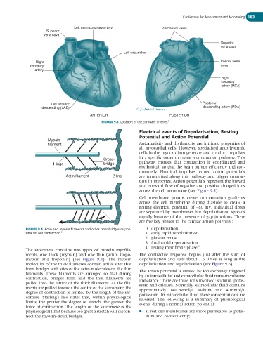
Left main coronary artery Pulmonary veins
Superior
vena cava
Superior
vena cava
Left circumflex
Right Inferior vena
coronary cava
artery
Right
coronary
artery (RCA)
Left anterior Posterior
descending (LAD) descending artery (PDA)
ANTERIOR POSTERIOR
FIGURE 9.3 Location of the coronary arteries. 5
Electrical events of Depolarisation, Resting
Potential and Action Potential
Myosin
filament Automaticity and rhythmicity are intrinsic properties of
all myocardial cells. However, specialised autorhythmic
cells in the myocardium generate and conduct impulses
in a specific order to create a conduction pathway. This
Cross-
Hinge bridge pathway ensures that contraction is coordinated and
rhythmical, so that the heart pumps efficiently and con-
tinuously. Electrical impulses termed action potentials
Actin filament Z line are transmitted along this pathway and trigger contrac-
tion in myocytes. Action potentials represent the inward
and outward flow of negative and positive charged ions
across the cell membrane (see Figure 9.5).
Cell membrane pumps create concentration gradients
across the cell membrane during diastole to create a
resting electrical potential of −80 mV. Individual fibres
are separated by membranes but depolarisation spreads
rapidly because of the presence of gap junctions. There
are five key phases to the cardiac action potential:
FIGURE 9.4 Actin and myosin filaments and other cross-bridges respon- 0. depolarisation
sible for cell contraction. 5 1. early rapid repolarisation
2. plateau phase
3. final rapid repolarisation
4. resting membrane phase. 8
The sarcomere contains two types of protein myofila-
ments, one thick (myosin) and one thin (actin, tropo- The contractile response begins just after the start of
myosin and troponin) (see Figure 9.4). The myosin depolarisation and lasts about 1.5 times as long as the
molecules of the thick filaments contain active sites that depolarisation and repolarisation (see Figure 9.6).
form bridges with sites of the actin molecules on the thin The action potential is created by ion exchange triggered
filaments. These filaments are arranged so that during by an intracellular and extracellular fluid trans-membrane
contraction, bridges form and the thin filaments are imbalance. There are three ions involved: sodium, potas-
pulled into the lattice of the thick filaments. As the fila- sium and calcium. Normally, extracellular fluid contains
ments are pulled towards the centre of the sarcomere, the approximately 140 mmol/L sodium and 4 mmol/L
degree of contraction is limited by the length of the sar- potassium. In intracellular fluid these concentrations are
comere. Starling’s law states that, within physiological reversed. The following is a summary of physiological
limits, the greater the degree of stretch, the greater the events during a normal action potential:
force of contraction. The length of the sarcomere is the
physiological limit because too great a stretch will discon- ● at rest cell membranes are more permeable to potas-
nect the myosin–actin bridges. sium and consequently;