
Page 211 - ACCCN's Critical Care Nursing
P. 211
188 P R I N C I P L E S A N D P R A C T I C E O F C R I T I C A L C A R E
impetus for blood through the pulmonary and systemic
vascular systems. The phases of the cardiac cycle are char- Increased
acterised by pressure changes within each of the heart contractility
chambers, resulting in blood flow from areas of high 125
pressure to areas of lower pressure.
During late ventricular diastole (rest), pressures are lowest 100 Normal
contractility
in the heart and blood returns passively to fill the atria.
This flow also moves into the ventricle through the open Ventricular stroke work (mmHg) 75
AV valves, producing 70–80% of ventricular filling. The Decreased
pulmonic and aortic valves are closed, preventing back- 50 contractility
flow from the pulmonary and systemic systems into the
ventricles. Depolarisation of the atria then occurs, some-
times referred to as atrial kick, stimulating atrial contrac- 25
tion and completing the remaining 20–30% of ventricular
filling.
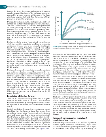
During ventricular systole (contraction), the atria relax 0 4 8 12 16
while the ventricles depolarise, resulting in ventricular Left ventricular end-diastolic filling pressure (LVEDP)
contraction. Pressure rises in the ventricles, resulting in FIGURE 9.10 The Frank-Starling curve. As left ventricular end-diastolic
the AV valves closing. When this occurs, all four cardiac pressure increases, so does ventricular stroke work. 5
valves are closed, blood volume is constant and contrac-
tion occurs (isovolumetric contraction). When the pres-
sure in the ventricles exceeds the pressure in the major According to this mechanism, within limits, the more
vessels the semilunar valves open. This occurs when pres- stretch on the cardiac muscle fibre before contraction, the
sure in the left ventricle reaches approximately 80 mmHg greater the strength of contraction. The ability to increase
and in the right ventricle approximately 27–30 mmHg. strength of contraction in response to increased stretch is
During the peak ejection phase, pressure in the left ven- because there is an optimal range of cross-bridges that
tricle and aorta reaches approximately 120 mmHg and in can be created between actin and myosin in the myocyte.
the right ventricle and pulmonary artery approximately Under this range, when venous return is poor, fewer
25–28 mmHg.
cross-bridges can be created. Above this range, when heart
During early ventricular diastole, the ventricles repolarise failure is present, the cross-bridges can become partially
and ventricular relaxation occurs. The pressure in the ven- disengaged, contraction is poor, and higher filling pres-
tricles falls until the pressures in the aorta and pulmonary sures are needed to achieve adequate contractile force.
artery are higher and blood pushes back against the semi- Ventricular contraction is also intrinsically influenced by
lunar valves. Shutting of these valves prevents backflow the size of the ventricle and the thickness of the ventricle
into the ventricles, and pressure in the ventricles declines wall. This mechanism is described by Laplace’s law, which
further. During ventricular contraction, the atria have states that the amount of tension generated in the wall
been filling passively, so the pressure in the atria rises to of the ventricle required to produce intraventricular pres-
higher than that in the ventricles and the AV valves open, sure depends on the size (radius and wall thickness) of
allowing blood flow to the ventricles. Any rise in heart the ventricle. As a result, in heart failure, when ventricu-
1
rate will shorten the resting period, which may impair lar thinning and dilation is present, more tension or
filling time and coronary artery flow as these arteries fill contractile force is required to create intraventricular pres-
during diastole. 1
sure and therefore cardiac output.
Regulation of Cardiac Output The heart’s ability to pump effectively is also influenced
The heart is a very effective pump and is able to adapt to by the pressure that is required to generate above end
meet the metabolic needs of the body. The activities of diastolic pressure to eject blood during systole. This addi-
the heart are regulated by two responsive systems: intrin- tional pressure is usually determined by how much resis-
sic regulation of contraction, and the autonomic nervous tance is present in the pulmonary artery and aorta, and
system. is in turn influenced by the peripheral vasculature. This
systemic vascular resistance, causing resistance to flow
Intrinsic regulation of contraction responds to the rate of
blood flow into the chambers. Blood flow into the heart known and measured as afterload, is in relation to the
depends on venous return from systemic and pulmonic left ventricle and is influenced by vascular tone and
veins and varies according to tissue metabolism, total disease.
blood volume and vasodilation. Venous return contrib-
utes to end-diastolic volume (preload) and pressure, Autonomic nervous system control and
which are both directly related to the force of contraction regulation of heart rate
in the next ventricular systole. The intrinsic capacity of Although the pacemaker cells of the heart are capable of
the heart to respond to changes in end-diastolic pressure intrinsic rhythm generation (automaticity), inputs from
can be represented by a number of length–tension curves the autonomic nervous system regulate heart rate changes
and the Frank-Starling mechanism (see Figure 9.10). in accordance with body needs by stimulating or