
Page 239 - ACCCN's Critical Care Nursing
P. 239
216 P R I N C I P L E S A N D P R A C T I C E O F C R I T I C A L C A R E
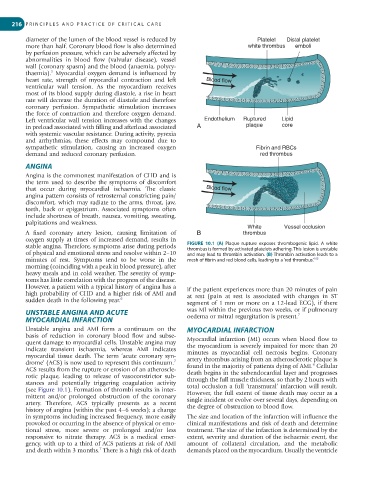
diameter of the lumen of the blood vessel is reduced by Platelet Distal platelet
more than half. Coronary blood flow is also determined white thrombus emboli
by perfusion pressure, which can be adversely affected by
abnormalities in blood flow (valvular disease), vessel
wall (coronary spasm) and the blood (anaemia, polycy-
5
thaemia). Myocardial oxygen demand is influenced by
heart rate, strength of myocardial contraction and left Blood flow
ventricular wall tension. As the myocardium receives
most of its blood supply during diastole, a rise in heart
rate will decrease the duration of diastole and therefore
coronary perfusion. Sympathetic stimulation increases
the force of contraction and therefore oxygen demand.
Lipid
Left ventricular wall tension increases with the changes Endothelium Ruptured Lipid
core
in preload associated with filling and afterload associated A plaque core
with systemic vascular resistance. During activity, pyrexia
and arrhythmias, these effects may compound due to
sympathetic stimulation, causing an increased oxygen Fibrin and RBCs
demand and reduced coronary perfusion. red thrombus
ANGINA
Angina is the commonest manifestation of CHD and is
the term used to describe the symptoms of discomfort
that occur during myocardial ischaemia. The classic Blood flow
angina pattern consists of retrosternal constricting pain/
discomfort, which may radiate to the arms, throat, jaw,
teeth, back or epigastrium. Associated symptoms often
include shortness of breath, nausea, vomiting, sweating,
palpitations and weakness.
White Vessel occlusion
A fixed coronary artery lesion, causing limitation of B thrombus
oxygen supply at times of increased demand, results in
stable angina. Therefore, symptoms arise during periods FIGURE 10.1 (A) Plaque rupture exposes thrombogenic lipid. A white
thrombus is formed by activated platelets adhering. This lesion is unstable
of physical and emotional stress and resolve within 2–10 and may lead to thrombin activation. (B) Thrombin activation leads to a
minutes of rest. Symptoms tend to be worse in the mesh of fibrin and red blood cells, leading to a ‘red thrombus’.
105
morning (coinciding with a peak in blood pressure), after
heavy meals and in cold weather. The severity of symp-
toms has little correlation with the progress of the disease.
However, a patient with a typical history of angina has a if the patient experiences more than 20 minutes of pain
high probability of CHD and a higher risk of AMI and at rest (pain at rest is associated with changes in ST
sudden death in the following year. 6 segment of 1 mm or more on a 12-lead ECG), if there
UNSTABLE ANGINA AND ACUTE was MI within the previous two weeks, or if pulmonary
7
oedema or mitral regurgitation is present.
MYOCARDIAL INFARCTION
Unstable angina and AMI form a continuum on the MYOCARDIAL INFARCTION
basis of reduction in coronary blood flow and subse- Myocardial infarction (MI) occurs when blood flow to
quent damage to myocardial cells. Unstable angina may the myocardium is severely impaired for more than 20
indicate transient ischaemia, whereas AMI indicates minutes as myocardial cell necrosis begins. Coronary
myocardial tissue death. The term ‘acute coronary syn- artery thrombus arising from an atherosclerotic plaque is
7
drome’ (ACS) is now used to represent this continuum. found in the majority of patients dying of AMI. Cellular
8
ACS results from the rupture or erosion of an atheroscle- death begins in the subendocardial layer and progresses
rotic plaque, leading to release of vasoconstrictor sub- through the full muscle thickness, so that by 2 hours with
stances and potentially triggering coagulation activity total occlusion a full ‘transmural’ infarction will result.
(see Figure 10.1). Formation of thrombi results in inter- However, the full extent of tissue death may occur as a
mittent and/or prolonged obstruction of the coronary single incident or evolve over several days, depending on
artery. Therefore, ACS typically presents as a recent the degree of obstruction to blood flow.
history of angina (within the past 4–6 weeks); a change
in symptoms including increased frequency, more easily The size and location of the infarction will influence the
provoked or occurring in the absence of physical or emo- clinical manifestations and risk of death and determine
tional stress, more severe or prolonged and/or less treatment. The size of the infarction is determined by the
responsive to nitrate therapy. ACS is a medical emer- extent, severity and duration of the ischaemic event, the
gency, with up to a third of ACS patients at risk of AMI amount of collateral circulation, and the metabolic
7
and death within 3 months. There is a high risk of death demands placed on the myocardium. Usually the ventricle