
Page 360 - ACCCN's Critical Care Nursing
P. 360
Respiratory Assessment and Monitoring 337
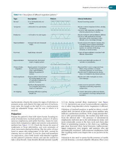
TABLE 13.1 Description of different respiration patterns 14
Type Description Pattern Clinical indication
Normal 12 to 20 breaths/min and Normal breathing pattern
regular
Tachypnea >24 breaths/min and shallow May be a normal response to fever, anxiety,
or exercise
Can occur with respiratory insufficiency,
alkalosis, pneumonia, or pleurisy
Bradypnea <10 breaths/min and regular May be normal in well-conditioned athletes
Can occur with medication-induced
depression of the respiratory centre,
diabetic coma, neurologic damage
Hyperventilation Increased rate and increased Usually occurs with extreme exercise, fear,
depth or anxiety. Causes of hyperventilation
include disorders of the central nervous
system, an overdose of the drug
salicylate, or severe anxiety.
Kussmaul Rapid, deep, laboured A type of hyperventilation associated with
diabetic ketoacidosis
Hypoventilation Decreased rate, decreased Usually associated with overdose of
depth, irregular pattern narcotics or anaesthetics
Cheyne-Stokes Regular pattern characterised May result form severe congestive heart
respiration by alternating periods of failure, drug overdose, increased
deep, rapid breathing intracranial pressure, or renal failure
followed by periods of May be noted in elderly persons during
apnoea sleep, not related to any disease process
Biot’s respiration Irregular pattern characterised May be seen with meningitis or severe
by varying depth and rate brain damage
of respirations followed by
periods of apnoea
Ataxic Significant disorganisation A more extreme expression of Biot’s
with irregular and varying respirations indicating respiratory
depths of respiration compromise
Air trapping Increasing difficulty in getting In chronic obstructive pulmonary disease,
breath out air is trapped in the lungs during forced
expiration
tracheostomy, observe the stoma for signs of infection or 3–5 cm during normal deep inspiration (see Figure
1
pressure areas; and observe the type and size of tracheos- 13.13). Asymmetry can occur in pneumothorax, pneumo-
tomy tube, the length at the hub if it is a tracheostomy nia or other lung disorders where inspiration is affected.
with an adjustable flange, and the way in which it is
secured. Palpation of tracheal position is useful to detect a medi-
astinal shift; deviation of the trachea from midline may
Palpation indicate a pulmonary problem. With a large pneumotho-
Palpate the patient’s chest with warm hands, focusing on: rax or after pneumonectomy, the trachea may shift away
28
areas of tenderness, tracheal position, presence of subcu- from the affected side. The presence of subcutaneous
taneous emphysema and tactile fremitus. Assess for sym- emphysema indicates air in the subcutaneous tissue and
metry (left compared to right) and anterior and posterior most commonly occurs in the face, neck and chest after
surfaces (see Figure 13.12). Check the thorax for areas of blunt or penetrating trauma to the chest (e.g. stabbing,
tenderness or bony deformities, and note symmetry of gun shot, fractured ribs); facial fractures; tracheostomy;
chest movement during breathing. Use the palm of your upper respiratory tract surgery; and patients who are
hand to assess skin temperature of the skin, noting for mechanically ventilated. Subcutaneous emphysema feels
clammy, hot or cold skin. To test for chest wall symmetry like crackling under your fingers due to air pockets in the
29
on inspiration, place both hands with thumbs together tissue.
on the patient’s posterior thorax and ask the patient to Palpation is also used to assess for the presence of tactile
take a deep breath. Your thumbs should separate equally (vocal) fremitus, a normal palpable vibration. Place your