
Page 362 - ACCCN's Critical Care Nursing
P. 362
Respiratory Assessment and Monitoring 339
no airflow through that area of the lung and also requires
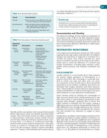
TABLE 13.2 Normal breath sounds 1 immediate treatment. 22,31
Sound Characteristics
Vesicular Heard over most of lung field; low pitch; soft Practice tip
and short exhalation, and long inhalation.
Respiratory rate is an early warning sign for respiratory distress.
Bronchovesicular Heard over main bronchus area and over If a patient has a high respiratory rate it can be a sign of hypoxia
upper right posterior lung field; medium
pitch; exhalation equals inhalation. as they attempt to compensate for a low PO 2 .
Bronchial Heard only over trachea; high pitch; loud
and long exhalation.
Documentation and Charting
Document the findings of your respiratory assessment in
the patient’s chart; if this is the first respiratory assess-
TABLE 13.3 Description of abnormal breath sounds 1 ment, describe the patient’s respiratory history carefully.
Any abnormal findings including abnormal sounds and
Abnormal their characteristics should be described to enable subse-
Sound Description Condition quent re-assessment. 30
Absent No airflow to Pneumothorax
breath particular Pneumonectomy RESPIRATORY MONITORING
sounds portion of lung Emphysematous blebs
Pleural effusion A thorough and comprehensive assessment, with accurate
Lung mass ongoing monitoring, enables early detection of condition
Massive atelectasis changes and assessment of responses to treatment for a
Complete airway
obstruction critically ill patient. This section describes the main
aspects of bedside respiratory monitoring and the instru-
Diminished Little airflow to Emphysema ments used to assess the efficiency of a patient’s gas
breath particular Pleural effusion
sounds portion of lung Pleurisy transfer mechanisms, including pulse oximetry, capno-
Atelectasis graphy, airway pressures and ventilator waveforms and
Pulmonary fibrosis loops.
Displaced Bronchial sounds Atelectasis with secretions
bronchial heard in Lung mass with exudates PULSE OXIMETRY
sounds peripheral lung Pneumonia
fields Pleural effusion A pulse oximeter is a non-invasive device that measures
Pulmonary oedema the arterial oxygen saturation of haemoglobin in a
patient’s blood flow. The technology is commonly stan-
Crackles Short, discrete Pulmonary oedema
(rales) popping or Pneumonia dard in critical care units and other acute care areas.
crackling sounds Pulmonary fibrosis It is important to note that the device does not provide
Atelectasis information on the patient’s ventilatory state, but it can
Bronchiectasis determine their oxygen saturation and detect hypoxae-
32
Rhonchi Coarse, rumbling, Pneumonia mia. This prompt non-invasive detection of hypoxaemia
low-pitched Asthma enables identification of clinical deterioration and more
sounds Bronchitis rapid treatment to avoid associated complications. 33
Bronchospasm
Pulse oximetry works by emitting two wavelengths of
Wheezes High-pitched, Asthma
squeaking, Bronchospasm light: red and infrared, from a diode (positioned on one
whistling sounds side of the probe) to a photodetector (positioned on the
opposite side) through a pulsatile flow of blood. The
Pleural Creaking, leathery, Pleural effusion
friction loud, dry, coarse Pleurisy signal emitted is measured over five pulses, causing a
rub sounds slight delay when monitoring. Oxygenated blood absorbs
light differently from deoxygenated blood; the oximeter
measures the amount of light absorbed by the vascular
bed and calculates the saturation of oxygen in those
Identify and become familiar with normal breath sounds capillaries.
before beginning to listen and identify abnormal breath
sounds. Abnormal breath sounds are either continuous Measurement of indirect arterial oxygen saturation of the
or discontinuous. Continuous sounds include wheezes peripheral circulation via pulse oximetry is referred to as
and rhonchi, while discontinuous sounds include crack- SpO 2 (the letter ‘p’ denotes peripheral) and is displayed
les (see Table 13.3). Stridor is an abnormal loud high- digitally on the monitor as a percentage, along with heart
pitched breath sound caused by obstruction in the upper rate and a plethysmographic waveform. Interpreting this
airways as a result of a foreign body, tissue swelling or waveform is essential in distinguishing a true oximetry
vocal cord; this emergent condition requires immediate signal from one displaying dampening or artefact (see
30
attention. Absent or diminished breath sounds indicate Figure 13.14). The probe is commonly sited on a finger,