
Page 440 - ACCCN's Critical Care Nursing
P. 440
Neurological Assessment and Monitoring 417
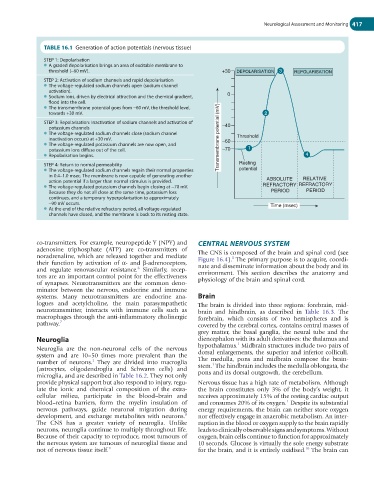
TABLE 16.1 Generation of action potentials (nervous tissue)
STEP 1: Depolarisation
● A graded depolarisation brings an area of excitable membrane to
threshold (−60 mV). +30 DEPOLARISATION 3 REPOLARISATION
STEP 2: Activation of sodium channels and rapid depolarisation
● The voltage-regulated sodium channels open (sodium channel
activation).
● Sodium ions, driven by electrical attraction and the chemical gradient, 0
flood into the cell.
● The transmembrane potential goes from −60 mV, the threshold level,
towards +30 mV. 2
STEP 3: Repolarisation: Inactivation of sodium channels and activation of
potassium channels −40
● The voltage-regulated sodium channels close (sodium channel Threshold
inactivation occurs) at +30 mV. Transmembrane potential (mV) −60
● The voltage-regulated potassium channels are now open, and
potassium ions diffuse out of the cell. −70 1
● Repolarisation begins. 4
STEP 4: Return to normal permeability Resting
● The voltage-regulated sodium channels regain their normal properties potential
in 0.4–1.0 msec. The membrane is now capable of generating another
action potential if a larger than normal stimulus is provided. ABSOLUTE RELATIVE
● The voltage-regulated potassium channels begin closing at −70 mV. REFRACTORY REFRACTORY
Because they do not all close at the same time, potassium loss PERIOD PERIOD
continues, and a temporary hyperpolarisation to approximately
−90 mV occurs. Time (msec)
● At the end of the relative refractory period, all voltage-regulated
channels have closed, and the membrane is back to its resting state.
co-transmitters. For example, neuropeptide Y (NPY) and CENTRAL NERVOUS SYSTEM
adenosine triphosphate (ATP) are co-transmitters of The CNS is composed of the brain and spinal cord (see
noradrenaline, which are released together and mediate Figure 16.4). The primary purpose is to acquire, coordi-
5
their function by activation of α- and β-adrenoceptors, nate and disseminate information about the body and its
and regulate renovascular resistance. Similarly, recep- environment. This section describes the anatomy and
6
tors are an important control point for the effectiveness physiology of the brain and spinal cord.
of synapses. Neurotransmitters are the common deno-
minator between the nervous, endocrine and immune
systems. Many neurotransmitters are endocrine ana- Brain
logues and acetylcholine, the main parasympathetic The brain is divided into three regions: forebrain, mid-
neurotransmitter, interacts with immune cells such as brain and hindbrain, as described in Table 16.3. The
macrophages through the anti-inflammatory cholinergic forebrain, which consists of two hemispheres and is
pathway. 7 covered by the cerebral cortex, contains central masses of
grey matter, the basal ganglia, the neural tube and the
Neuroglia diencephalon with its adult derivatives: the thalamus and
1
Neuroglia are the non-neuronal cells of the nervous hypothalamus. Midbrain structures include two pairs of
system and are 10–50 times more prevalent than the dorsal enlargements, the superior and inferior colliculi.
number of neurons. They are divided into macroglia The medulla, pons and midbrain compose the brain-
1
1
(astrocytes, oligodendroglia and Schwann cells) and stem. The hindbrain includes the medulla oblongata, the
microglia, and are described in Table 16.2. They not only pons and its dorsal outgrowth, the cerebellum.
provide physical support but also respond to injury, regu- Nervous tissue has a high rate of metabolism. Although
late the ionic and chemical composition of the extra- the brain constitutes only 3% of the body’s weight, it
cellular milieu, participate in the blood–brain and receives approximately 15% of the resting cardiac output
1
blood–retina barriers, form the myelin insulation of and consumes 20% of its oxygen. Despite its substantial
nervous pathways, guide neuronal migration during energy requirements, the brain can neither store oxygen
8
development, and exchange metabolites with neurons. nor effectively engage in anaerobic metabolism. An inter-
The CNS has a greater variety of neuroglia. Unlike ruption in the blood or oxygen supply to the brain rapidly
neurons, neuroglia continue to multiply throughout life. leads to clinically observable signs and symptoms. Without
Because of their capacity to reproduce, most tumours of oxygen, brain cells continue to function for approximately
the nervous system are tumours of neuroglial tissue and 10 seconds. Glucose is virtually the sole energy substrate
10
not of nervous tissue itself. 9 for the brain, and it is entirely oxidised. The brain can