
Page 445 - ACCCN's Critical Care Nursing
P. 445
422 P R I N C I P L E S A N D P R A C T I C E O F C R I T I C A L C A R E
TABLE 16.3, Continued
Division Description Functions
Medulla oblongata Between the pons and the spinal The medulla oblongata contains motor fibres from the brain to
cord. the spinal cord and sensory fibres from the spinal cord to the
brain. Most of these fibres cross at this level. Cranial nerves
IX–XII connect to the brain in the medulla, which has centres
for control of vital functions, such as respiration and the heart
rate.
Cerebellum Below the posterior portion of the Coordinates voluntary muscles; maintains balance and muscle
cerebellum. Divided into two tone; has both excitatory and inhibitory actions. It also
hemispheres. controls fine movement, balance, position sense and
integration of sensory input.
Central Primary sensory
Primary motor cortex sulcus cortex
(precentral gyrus) (postcentral gyrus)
Somatic motor association
area (premotor cortex) PARIETAL LOBE
Parieto-occipital sulcus
FRONTAL LOBE Somatic sensory
association area
Prefrontal cortex Visual association area
OCCIPITAL LOBE
Gustatory cortex
Visual cortex
Insula Auditory association area
Lateral sulcus
Auditory cortex
Olfactory cortex
TEMPORAL LOBE
Frontal eye field
Speech centre A
4
6
1
General interpretive
area 40 39
44 41 42
16
17
Prefrontal
cortex B C
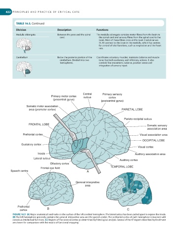
FIGURE 16.5 (A) Major anatomical landmarks on the surface of the left cerebral hemisphere. The lateral sulcus has been pulled apart to expose the insula.
(B) The left hemisphere generally contains the general interpretive area and the speech centre. The prefrontal cortex of each hemisphere is involved with
conscious intellectual functions. (C) Regions of the cerebral cortex as determined by histological analysis. Several of the 47 regions described by Brodmann
are shown for comparison with the results of functional mapping. 1