
Page 738 - ACCCN's Critical Care Nursing
P. 738
Pregnancy and Postpartum Considerations 715
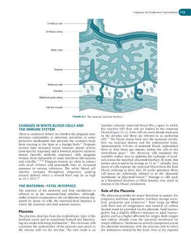
Umbilical vein
Umbilical artery
Main villus
Septum
Maternal vein
Decidua
Maternal spiral artery
Uterine muscle
FIGURE 26.1 The maternal–placental interface.
11
CHANGES IN WHITE BLOOD CELLS AND interface whereby maternal blood fills a space in which
THE IMMUNE SYSTEM the nutritive villi float and are bathed in the maternal
There is continued debate on whether the pregnant state blood (Figure 26.1). A few villi are more deeply anchored
increases vulnerability to infection, secondary to some in the decidua and these are referred to as anchoring
61
protective mechanism that prevents the woman’s body villi. The blood drains back into the maternal circula-
17
from reacting to the fetus as a foreign body. Pregnant tion via maternal sinuses and the endometrial veins.
women have increased innate immune system activity Approximately 150 mL of maternal blood, replenished
(non-specific response) and a lowered adaptive immune three to four times per minute, bathes the villi in the
61
system (specific antibody response), with pregnant intervillous space. The chorionic villi maximise the
women more vulnerable to some infections like malaria available surface area to optimise the exchange of prod-
and varicella. 17,59,60 Pregnant women are often in contact ucts across the maternal–placental interface. By term, this
2 62
with small children and potentially have an increased surface area is said to be as large as 13 m . Initially, four
exposure to various infections. The white blood cell layers of cells separate the maternal blood from the fetal
number increases throughout pregnancy, peaking blood, reducing to three after 20 weeks’ gestation; these
around delivery when a normal level may be as high cell layers are collectively referred to as the ‘placental
63
9
as 25 × 10 /L. 46 membrane’ or ‘placental barrier’. Damage to villi, such
as a threatened abortion or blunt trauma, may result in
THE MATERNAL–FETAL INTERFACE mixing of the blood circulations.
The junction of the maternal and fetal circulations is Role of the Placenta
referred to as the maternal–fetal interface. Although, The placenta provides six major functions to sustain the
under normal circumstances the circulations remain sep- pregnancy and fetus: respiration, nutrition, storage, excre-
arated by layers of cells, the maternal–fetal interface is tion, protection and endocrine. Fetal lungs are filled
61
where the maternal and fetal systems interact.
with fluid and all oxygenation and removal of carbon
dioxide must be provided via the placenta. Fetal haemo-
Placenta globin has a slightly different structure to adult haemo-
The placenta develops from the trophoblastic layer of the globin and has a higher affininity for oxygen. Both oxygen
fertilised ovum and is completely formed and function- and carbon dioxide cross the placental membrane by
61
ing ten weeks following fertilisation. The chorionic villi simple diffusion. Nutrients are actively transported across
constitute the undersurface of the placenta and attach to the placental membrane, with the placenta able to select
the uterine wall via the decidua. The end result is an the substances needed by the fetus, even at the expense