


Page 156 - Concise Pathology for Exam Preparation ( PDFDrive )
P. 156
6  Neoplasia  141


             •  Angiogenesis is required not only for tumour growth, but also for metastasis. Without
               access to the vasculature, the tumour cells cannot spread to distant sites.
             •  Sprouting of new vessels from already-existing ones is called neoangiogenesis; whereas,
               vasculogenesis  occurs  by  recruitment  of  endothelial  cell  precursors.  Tumour  blood
               vessels differ from the normal vasculature by being leaky and unorganized.
             •  The pro-angiogenic factors are produced by tumour cells or derived from inflammatory
               cells (eg, macrophages) that infiltrate tumours. Hypoxia activates transcription factor
               Hypoxia-inducible factor (HIF)-1a which in turn activates pro-angiogenic factors like
               VEGF and bFGF. Other promoters of angiogenesis include angiopoietins 1 and 2. In-
               hibitors of VEGF are being used for the treatment of advanced cancers. These are not
               curative but can prolong survival.
             •  Under normal circumstances, p53 increases expression of anti-angiogenic factors like thrombo-
               spondin-1, angiostatin, tumstatin and endostatin. Loss of p53, therefore, allows angiogenesis.

             Q. Describe the mechanism and biology of invasion and metastasis.
             Ans. In the early stage of tumourigenesis, the cells are not invasive and metastatic. Progres-
             sive genetic aberrations are associated with the appearance of new clones with invasiveness
             and metastatic ability. Highly metastatic cells often acquire alterations in more genes than
             nonmetastatic cells. Potentially important genes associated with epithelial metastasis include
             twist-related protein (TWIST) and the zinc-finger group gene SNAIL; these genes encode
             transcription  factors  which  increase  epithelial-mesenchymal  interactions.  The  following
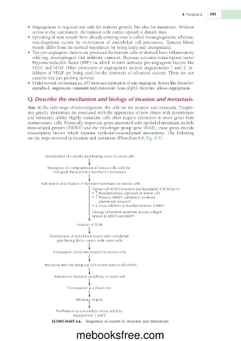
             are the steps involved in invasion and metastasis (Flowchart 6.6; Fig. 6.5):


                     Development of a rapidly proliferating clone of cancer cells

                      Emergence of a subpopulation of tumour cells with the
                        biological characteristics essential for metastases

                   Adhesion to and invasion of basement membrane by tumour cells
                                         Tumour cell–ECM interactions and degradation of ECM due to:
                                         • ↑ Metalloproteinase expression on tumour cells
                                         • ↑ Proteases (MMPs, cathepsin D, urokinase
                                               plasminogen activator)
                                         • ↓ Tissue inhibitors of metalloproteinases (TIMPs)
                                         Cleavage of basement membrane proteins collagen
                                         laminin by MMP2 and MMP9
                                  Invasion of ECM

                      Development of leaky blood vessels with endothelial
                          gaps having direct contact with cancer cells


                        Intravasation (entry into vessels) by tumour cells

                    Interaction with host lymphoid cells to form tumour cell emboli

                        Adhesion to basement membrane of vessel wall

                              Extravasation at a distant site

                                  Metastatic deposit


                         Proliferation as a secondary colony aided by
                                 angiopoietins 1 and 2
                        FLOWCHART 6.6.  Sequence of events in invasion and metastasis.


                                  mebooksfree.com

