
Page 202 - Concise Pathology for Exam Preparation ( PDFDrive )
P. 202
7 Infections 187
• When pigs ingest the proglottids or eggs, the eggs hatch, penetrate their intestinal wall,
and spread to skeletal muscle, especially the neck, tongue and trunk. There, the larvae
mature into encysted cysticerci over 2–3 months.
• The cysticerci suppress the host inflammatory response and survive in tissues for
months to years. The life cycle is completed when humans ingest inadequately cooked
pork that contains viable cysticerci or eggs.
• The larvae hatch, penetrate the gut wall, disseminate haematogenously, and encyst in
many organs. The egg-containing faeces usually contaminate water supplies in endemic
areas. If this water is used to irrigate fruits and vegetables, eggs are ingested with the
contaminated food. Thus, people who have never visited endemic countries can also
develop infection.
• Autoinfection involves the retrograde transmission of proglottids from the intestines
into the stomach with subsequent release of T. solium eggs into the gut.
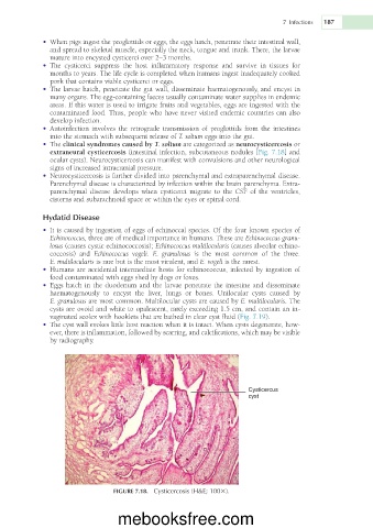
• The clinical syndromes caused by T. solium are categorized as neurocysticercosis or
extraneural cysticercosis (intestinal infection, subcutaneous nodules [Fig. 7.18] and
ocular cysts). Neurocysticercosis can manifest with convulsions and other neurological
signs of increased intracranial pressure.
• Neurocysticercosis is further divided into parenchymal and extraparenchymal disease.
Parenchymal disease is characterized by infection within the brain parenchyma. Extra-
parenchymal disease develops when cysticerci migrate to the CSF of the ventricles,
cisterns and subarachnoid space or within the eyes or spinal cord.
Hydatid Disease
• It is caused by ingestion of eggs of echinoccal species. Of the four known species of
Echinococcus, three are of medical importance in humans. These are Echinococcus granu-
losus (causes cystic echinococcosis); Echinococcus multilocularis (causes alveolar echino-
coccosis) and Echinococcus vogeli. E. granulosus is the most common of the three.
E. multilocularis is rare but is the most virulent, and E. vogeli is the rarest.
• Humans are accidental intermediate hosts for echinococcus, infected by ingestion of
food contaminated with eggs shed by dogs or foxes.
• Eggs hatch in the duodenum and the larvae penetrate the intestine and disseminate
haematogenously to encyst the liver, lungs or bones. Unilocular cysts caused by
E. granulosus are most common. Multilocular cysts are caused by E. multilocularis. The
cysts are ovoid and white to opalescent, rarely exceeding 1.5 cm, and contain an in-
vaginated scolex with hooklets that are bathed in clear cyst fluid (Fig. 7.19).
• The cyst wall evokes little host reaction when it is intact. When cysts degenerate, how-
ever, there is inflammation, followed by scarring, and calcifications, which may be visible
by radiography.
Cysticercus
cyst
FIGURE 7.18. Cysticercosis (H&E; 1003).
mebooksfree.com