
Page 201 - Concise Pathology for Exam Preparation ( PDFDrive )
P. 201
186 SECTION I General Pathology
Mucosa
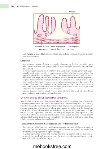
Muscularis mucosae Flask shaped ulcer Lamina propria
FIGURE 7.17. A flask-shaped amoebic ulcer.
pasty (anchovy sauce-like) material. These may undergo secondary bacterial infection
causing suppuration.
Diagnosis
• Asymptomatic human infections are usually diagnosed by finding cysts shed in the
stool. Various sedimentation procedures have been developed to recover the cysts from
faecal matter.
• In symptomatic infections, the motile form (trophozoite) can often be seen in fresh faeces.
• Amoebic trophozoites can also be demonstrated in histopathology sections, where they
appear as spherical or oval-shaped bodies (15–20 microns in diameter) with a thin cell
membrane and single nucleus with prominent nuclear border and central karyosome.
Trophozoites resemble macrophages because of a comparable size and presence of mul-
tiple vacuoles; the parasite, however, has a smaller nucleus with a large karyosome. The
PAS procedure stains the cytoplasm of the trophozoite red. The organism appears black
when stained with Heidenhain’s iron haematoxylin method. Presence of trophozoites
containing RBCs is indicative of tissue invasion.
• Serology becomes positive about 2 weeks after infection. The levels of antibody are
much higher in individuals with liver abscesses.
Q. Write briefly about helminthic infections.
Ans. The helminths are worm-like, multicellular parasites. They undergo sexual reproduc-
tion in the definitive host and asexual multiplication in an intermediary host. The clinically
important helminths are classified according to their physical characteristics, internal mor-
phology (appearance of their egg, larval and adult stages), as well as, and the host/vector
they inhabit. Flukes (Trematodes) are leaf-shaped flatworms with prominent oral and
ventral suckers. Tapeworms (Cestodes) are elongated, segmented, hermaphroditic flat-
worms that inhabit the intestinal lumen. Larval forms, which are cystic or solid, inhabit
extraintestinal tissues. Roundworms (Nematodes) are bisexual, cylindrical worms. They
inhabit intestinal and extraintestinal sites.
Tapeworms (Cestodes): Cysticercosis and Hydatid Disease
• Taenia solium and Echinococcus granulosus are cestodes (tapeworms) that cause cysticer-
cosis and hydatid infections, respectively. Both diseases are caused by larvae that
develop following ingestion of tapeworm eggs.
• T. solium tapeworms consist of a head (scolex) that has suckers and hooklets that at-
tach to the intestinal wall, a neck and many flat segments called proglottids that contain
male and female reproductive organs.
mebooksfree.com