
Page 199 - Concise Pathology for Exam Preparation ( PDFDrive )
P. 199
184 SECTION I General Pathology
The types of leishmaniasis are depicted in Table 7.6.
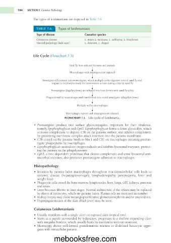
TABLE 7.6. Types of leishmaniasis
Type of disease Causative species
Cutaneous disease L. major, L. mexicana, L. aethiopica, L. braziliensis
Visceral pathology (kala azar) L. donovani, L. chagasi
Life Cycle (Flowchart 7.5)
Sand fly bites infected humans and animals
Macrophages with amastigotes are ingested
Amastigotes differentiate into promastigotes, which multiply in the digestive tract of sand fly and
migrate to its pharynx ready for transmission to host during a bite by sand fly
Promastigotes (flagellate forms) are released into host dermis with sand fly saliva
Phagocytosed by macrophages and transformed into round amastigotes (aflagellate forms)
Multiply within macrophages
Macrophages rupture and amastigotes are released
FLOWCHART 7.5. Life cycle of Leishmania.
• Promastigotes produce two surface glycoconjugates, important for their virulence,
namely, lipophosphoglycan and Gp63. Lipophosphoglycan forms a dense glycocalyx, which
activates complement to deposit C3b on the parasite surface, and inhibits complement
by preventing membrane complex attack insertion into the parasite membrane.
• C3b coated on the parasite binds to Mac1 and CR1 on macrophages initiating promas-
tigote phagocytosis by macrophages.
• Lipophosphoglycan neutralizes oxygen radicals and inhibits lysosomal enzymes, protect-
ing the parasite in the phagolysosome.
• Gp63, a zinc-dependent proteinase that cleaves complement and some lysosomal anti-
microbial enzymes; also promotes promastigote adhesion to macrophages.
Histopathology
• Invasion by parasite-laden macrophages throughout reticuloendothelial cells leads to
systemic disease (hepatosplenomegaly, lymphadenopathy, pancytopenia, fever and
weight loss).
• Phagocytic cells crowd the bone marrow, lymph nodes, liver, lungs, GIT, kidneys, pancreas
and testes.
• Liver becomes fibrotic in later stages. Normal architecture of the spleen may be replaced
by sheets of histiocytes, which are parasite laden. Plasma cells are increased in number.
• Kidney biopsy may show mesangioproliferative glomerulonephritis and/or amyloidosis.
• Hyperpigmentation of the skin (black fever) may be seen.
Cutaneous Leishmaniasis
• Usually manifests with a single ulcer on exposed skin (tropical sore).
• Starts as a papule surrounded by induration, progresses to a shallow expanding ulcer
with irregular borders, which usually heals by involution without treatment.
• Microscopy shows well-formed granulomatous reaction or ill-defined histiocytic aggre-
gates with intracellular parasite.
mebooksfree.com