
Page 200 - Concise Pathology for Exam Preparation ( PDFDrive )
P. 200
7 Infections 185
Mucocutaneous Leishmaniasis
• Moist, ulcerating and nonulcerating lesions arise in larynx, nasal septum and vulva after
the skin lesion has healed.
• Microscopy shows histiocytes, lymphocytes, plasma cells and occasionally granuloma-
tous reaction.
Diffuse Cutaneous Leishmaniasis in Anergic Patients
Starts as a single nodule and spreads to the whole body as bizarre nodular lesions (resemble
keloids and verrucae; they may sometimes be mistaken for the nodules of lepromatous leprosy).
Amoebiasis
• Amoebiasis is caused by Entamoeba histolytica, a protozoan parasite, which spreads by
faecal-oral transmission. Amoeba proteins involved in tissue invasion include
• Cysteine proteases, which lyse proteins of extracellular matrix.
• Lectins on parasite surface that bind to carbohydrates on colonic epithelium and RBCs.
• Channel forming proteins that contains an ameba pore that makes pores in plasma
membrane and lyses it.
• It is important to distinguish the E. histolytica cyst from the cysts of nonpathogenic
intestinal protozoa such as Entamoeba coli.
• E. histolytica cysts have a maximum of four nuclei, while the commensal E. coli cyst
has up to 8 nuclei.
• In E. histolytica, the endosome is centrally located in the nucleus, while it is usually
eccentric in E. coli.
• Chromatoidal bodies in E. histolytica cysts are rounded, while they are jagged in
E. coli.
• Virulent strains of E. histolytica show ingested RBCs, whereas nonvirulent strains do not.
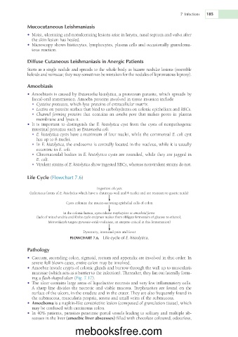
Life Cycle (Flowchart 7.6)
Ingestion of cysts
(Infectious forms of E. histolytica which have a chitinous wall and 4 nuclei and are resistant to gastric acids)
Cysts colonize the mucin-secreting epithelial cells of colon
In the colonic lumen, cysts release trophozoites or amoeboid forms
(lack of mitochondria and Krebs cycle enzymes makes them obligate fermenters of glucose to ethanol;
Metronidazole targets pyruvate–oxido-reductase, an enzyme critical in this fermentation)
Dysentery, intestinal pain and fever
FLOWCHART 7.6. Life cycle of E. histolytica.
Pathology
• Caecum, ascending colon, sigmoid, rectum and appendix are involved in that order. In
severe full blown cases, entire colon may be involved.
• Amoebae invade crypts of colonic glands and burrow through the wall up to muscularis
mucosae (which acts as a barrier to the infection). Thereafter, they fan out laterally form-
ing a flask-shaped ulcer (Fig. 7.17).
• The ulcer contains large areas of liquefactive necrosis and very few inflammatory cells.
A sharp line divides the necrotic and viable mucosa. Trophozoites are found on the
surface of the ulcers, in the exudate and in the crater. They are also frequently found in
the submucosa, muscularis propria, serosa and small veins of the submucosa.
• Amoeboma is a napkin-like constrictive lesion (composed of granulation tissue), which
may be confused with carcinoma colon.
• In 40% patients, parasites penetrate portal vessels leading to solitary and multiple ab-
scesses in the liver (amoebic liver abscesses) filled with chocolate coloured, odourless,
mebooksfree.com