
Page 304 - Concise Pathology for Exam Preparation ( PDFDrive )
P. 304
12 Haematology 289
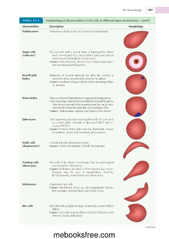
TABLE 12.4. Morphological abnormalities of red cells in different types of anaemias—cont’d
Abnormalities Description Morphology
Poikilocytosis Variation in shape of the cell as seen in thalassaemias
Target cells Flat red cells with a central mass of haemoglobin (dense
(codocytes) area), surrounded by a ring of pallor (pale area) and an
outer ring of haemoglobin (dense area)
Causes: Iron deficiency, chronic liver disease, hyposplen-
ism and haemoglobinopathies
Howell–Jolly Remnants of nuclear material left after the nucleus is
bodies extruded, these are normally removed by spleen
Causes: Nonfunctioning or absent spleen and megaloblas-
tic anaemia
Heinz bodies These are formed from denatured aggregated haemoglobin.
Cells containing Heinz bodies are normally removed by spleen.
They do not stain with Romanowski stains but can be dem-
onstrated by Supravital stains like new methylene blue.
Causes: Thalassaemia, asplenia and chronic liver disease
Spherocytes Dark appearing (densely haemoglobinized) red cells with
no central pallor (normal or decreased MCV and in-
creased MCHC)
Causes: Immune haemolytic anaemia, haemolytic disease
of newborn, burns and hereditary spherocytosis
Sickle cells Curved red cells with pointed ends
(drepanocytes) Causes: Sickle-cell anaemia, HbS-b Thalassaemia
Teardrop cells Red cells in the shape of teardrops; they are pathological
(dacrocytes) and should be followed up
Causes: Infiltrative disorders of bone marrow (eg, myelo-
fibrosis); may be seen in megaloblastic anaemia,
b Thalassaemia, renal failure and tuberculosis
Schistocytes Fragmented red cells
Causes: Mechanical stress, eg, microangiopathic haemo-
lytic anaemia, thermal injury and severe burns
Bite cells Red cells with peripheral single or multiple arcuate defects
(bites)
Causes: Red cells enzyme defects (G-6-PD deficiency and
Pyruvate kinase deficiency)
Continued
mebooksfree.com