


Page 42 - Concise Pathology for Exam Preparation ( PDFDrive )
P. 42
1  Cell Injury and Cell Death  27


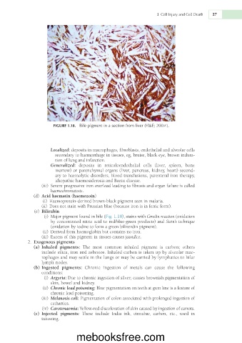
                        FIGURE 1.18.  Bile pigment in a section from liver (H&E; 2003).


                       Localized: deposits in macrophages, fibroblasts, endothelial and alveolar cells
                         secondary to haemorrhage in tissues, eg, bruise, black eye, brown indura-
                         tion of lung and infarction.
                       Generalized:  deposits  in  reticuloendothelial  cells  (liver,  spleen,  bone
                         marrow) or parenchymal organs (liver, pancreas, kidney, heart) second-
                         ary to haemolytic disorders, blood transfusions, parenteral iron therapy,
                         idiopathic haemosiderosis and Bantu disease.
                     (iii)  Severe progressive iron overload leading to fibrosis and organ failure is called
                       haemochromatosis.
                  (d)  Acid haematin (haemozoin)
                    (i)  Haemoprotein-derived brown-black pigment seen in malaria.
                     (ii)  Does not stain with Prussian blue (because iron is in ferric form).
                 (e)  Bilirubin
                     (i)  Major pigment found in bile (Fig. 1.18), stains with Gmelin reaction (oxidation
                       by concentrated nitric acid to red/blue-green products) and Stein’s technique
                       (oxidation by iodine to form a green biliverdin pigment).
                    (ii)  Derived from haemoglobin but contains no iron.
                     (iii)  Excess of this pigment in tissues causes jaundice.
               2.  Exogenous pigments
                  (a)  Inhaled  pigments:  The  most  common  inhaled  pigment  is  carbon;  others
                   include silica, iron and asbestos. Inhaled carbon is taken up by alveolar mac-
                   rophages and may settle in the lungs or may be carried by lymphatics to hilar
                   lymph nodes.
                  (b) Ingested  pigments:  Chronic  ingestion  of  metals  can  cause  the  following
                   conditions:
                     (i)  Argyria: Due to chronic ingestion of silver; causes brownish pigmentation of
                       skin, bowel and kidney.
                    (ii)  Chronic lead poisoning: Blue pigmentation on teeth at gum line is a feature of
                       chronic lead poisoning.
                     (iii)  Melanosis coli: Pigmentation of colon associated with prolonged ingestion of
                       cathartics.
                    (iv)  Carotenaemia: Yellow-red discoloration of skin caused by ingestion of carrots.
                 (c)  Injected  pigments:  These  include  India  ink,  cinnabar,  carbon,  etc.,  used  in
                   tattooing.


                                  mebooksfree.com

