
Page 488 - Concise Pathology for Exam Preparation ( PDFDrive )
P. 488
16 Diseases of the Kidney and Lower Urinary Tract 473
Thickened
basement
membrane
Hypercellular
glomerular
Accentuation of
lobulation
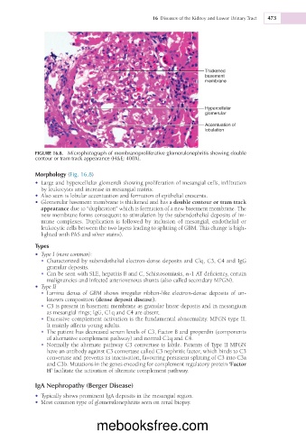
FIGURE 16.8. Microphotograph of membranoproliferative glomerulonephritis showing double
contour or tram track appearance (H&E; 400X).
Morphology (Fig. 16.8)
• Large and hypercellular glomeruli showing proliferation of mesangial cells, infiltration
by leukocytes and increase in mesangial matrix.
• Also seen is lobular accentuation and formation of epithelial crescents.
• Glomerular basement membrane is thickened and has a double contour or tram track
appearance due to “duplication” which is formation of a new basement membrane. The
new membrane forms consequent to stimulation by the subendothelial deposits of im-
mune complexes. Duplication is followed by inclusion of mesangial, endothelial or
leukocytic cells between the two layers leading to splitting of GBM. This change is high-
lighted with PAS and silver stains).
Types
• Type I (more common):
• Characterized by subendothelial electron-dense deposits and Clq, C3, C4 and IgG
granular deposits.
• Can be seen with SLE, hepatitis B and C, Schistosomiasis, a-1 AT deficiency, certain
malignancies and infected arteriovenous shunts (also called secondary MPGN).
• Type II
• Lamina densa of GBM shows irregular ribbon-like electron-dense deposits of un-
known composition (dense deposit disease).
• C3 is present in basement membrane as granular linear deposits and in mesangium
as mesangial rings; IgG, C1q and C4 are absent.
• Excessive complement activation is the fundamental abnormality. MPGN type II.
It mainly affects young adults.
• The patient has decreased serum levels of C3, Factor B and properdin (components
of alternative complement pathway) and normal C1q and C4.
• Normally the alternate pathway C3 convertase is labile. Patients of Type II MPGN
have an antibody against C3 convertase called C3 nephritic factor, which binds to C3
convertase and prevents its inactivation, favouring persistent splitting of C3 into C3a
and C3b. Mutations in the genes encoding for complement regulatory protein ‘Factor
H’ facilitate the activation of alternate complement pathway.
IgA Nephropathy (Berger Disease)
• Typically shows prominent IgA deposits in the mesangial region.
• Most common type of glomerulonephritis seen on renal biopsy.
mebooksfree.com