
Page 586 - Concise Pathology for Exam Preparation ( PDFDrive )
P. 586
21 Musculoskeletal System 571
that are associated with proteins like osteopontin. The inorganic part is constituted
by hydroxyapatite which serves as a reservoir for calcium.
Q. Define and classify osteomyelitis.
Ans. Osteomyelitis is defined as infection of the bone (osteo) and marrow (myelo) by
bacteria, viruses or fungi.
Classification
1. Pyogenic (bacterial) osteomyelitis:
• Most frequently targets children and young adults.
• Occurs due to haematogenous spread; extension from a contiguous site of infection,
eg, cellulitis or direct implantation.
• Common causative organisms include Staphylococcus aureus (in 80–90% cases)
followed by E. coli, Pseudomonas, Klebsiella, Haemophilus influenzae and Salmonella.
Mixed infections are also seen.
• The most frequent sites of involvement are the areas of rapid growth (distal femur,
proximal tibia, proximal humerus and distal radius).
• Location of infection is influenced by the vascular circulation. The slowing or sludg-
ing of blood flow as the vessels make sharp angles at the metaphyses predisposes the
vessels to thrombosis and the bone itself to localized necrosis and bacterial seeding.
• In the presence of bacterial infection elsewhere, a site of thrombosis acts as a nidus
for bacterial growth and development of osteomyelitis.
• Trauma is an important predisposing factor for osteomyelitis because it aids in
venostasis and thrombosis.
Stages of infection:
• Acute (develops over days and weeks)
• Chronic (develops over weeks to months; may persist for years)
Pathology:
Acute followed by chronic inflammatory cells are seen surrounding fragments of dead
bone. Occasionally foreign body giant cell reaction may be seen (Fig 21.3). The dead
bone shows empty lacunae without osteocytes.
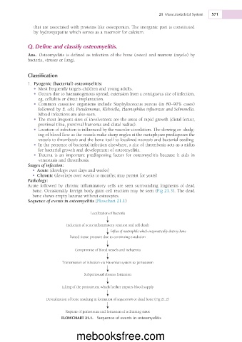
Sequence of events in osteomyelitis (Flowchart 21.1)
Localization of bacteria
Induction of acute inflammatory reaction and cell death
Influx of neutrophils which enzymatically destroy bone
Raised tissue pressure due to continuing exudation
Compromise of blood vessels and ischaemia
Transmission of infection via Haversian system to periosteum
Subperiosteal abscess formation
Lifting of the periosteum, which further impairs blood supply
Devitalization of bone resulting in formation of sequestrum or dead bone (Fig 21.2)
Rupture of periosteum and formation of a draining sinus
FLOWCHART 21.1. Sequence of events in osteomyelitis
mebooksfree.com