
Page 590 - Concise Pathology for Exam Preparation ( PDFDrive )
P. 590
21 Musculoskeletal System 575
Gross Morphology
• Sessile, round to oval and bosselated
• Project from subperiosteal/endosteal surface of cortex
• Multiple osteomas, may present with intestinal polyposis and soft tissue tumours
(Gardner syndrome)
Microscopy
Composed of dense and mature lamellar bone.
Osteoid Osteoma
Skeletal Distribution
• Long bones (femur and tibia)
• Usually intracortical; less frequently arise from medullary cavity
Clinical Features
• Common in the age group between 10 and 30 years.
• Presents with intense pain which increases during night and is relieved by aspirin
(pain is attributed to excessive PGE2 produced by proliferating osteoblasts). It may be
accompanied by localized swelling and tenderness.
X-ray
Shows a central nidus smaller than 1.5 cm that is surrounded by sclerotic bone. The nidus
may be difficult to see on plain X-ray. CT is modality of choice to identify it.
Gross Morphology
Appears as a well-defined, round-to-oval mass of gritty tissue with a size less than 2 cm
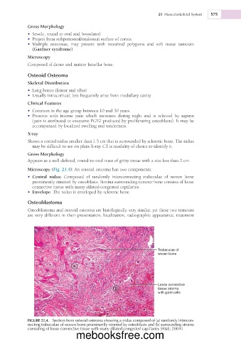
Microscopy (Fig. 21.4): An osteoid osteoma has two components:
• Central nidus: Composed of randomly interconnecting trabeculae of woven bone
prominently rimmed by osteoblasts. Stroma surrounding tumour bone consists of loose
connective tissue with many dilated-congested capillaries.
• Envelope: The nidus is enveloped by sclerotic bone.
Osteoblastoma
Osteoblastoma and osteoid osteoma are histologically very similar, yet these two tumours
are very different in their presentation, localization, radiographic appearance, treatment
Trabeculae of
woven bone
a
Loose connective
b tissue stroma
with giant cells
FIGURE 21.4. Section from osteoid osteoma showing a nidus composed of (a) randomly intercon-
necting trabeculae of woven bone prominently rimmed by osteoblasts and (b) surrounding stroma
consisting of loose connective tissue with many dilated-congested capillaries (H&E; 2003)
mebooksfree.com