
Page 589 - Concise Pathology for Exam Preparation ( PDFDrive )
P. 589
574 SECTION II Diseases of Organ Systems
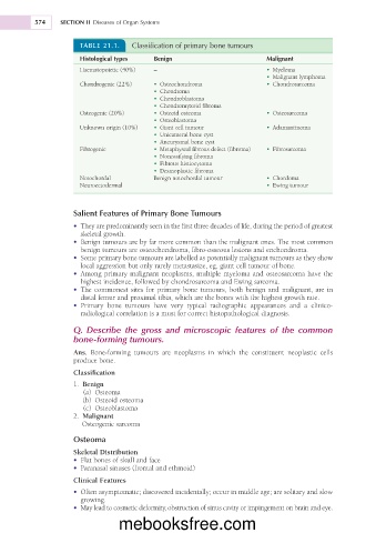
TABLE 21.1. Classification of primary bone tumours
Histological types Benign Malignant
Haematopoietic (40%) – • Myeloma
• Malignant lymphoma
Chondrogenic (22%) • Osteochondroma • Chondrosarcoma
• Chondroma
• Chondroblastoma
• Chondromyxoid fibroma
Osteogenic (20%) • Osteoid osteoma • Osteosarcoma
• Osteoblastoma
Unknown origin (10%) • Giant cell tumour • Adamantinoma
• Unicameral bone cyst
• Aneurysmal bone cyst
Fibrogenic • Metaphyseal fibrous defect (fibroma) • Fibrosarcoma
• Nonossifying fibroma
• Fibrous histiocytoma
• Desmoplastic fibroma
Notochordal Benign notochordal tumour • Chordoma
Neuroectodermal • Ewing tumour
Salient Features of Primary Bone Tumours
• They are predominantly seen in the first three decades of life, during the period of greatest
skeletal growth.
• Benign tumours are by far more common than the malignant ones. The most common
benign tumours are osteochondroma, fibro-osseous lesions and enchondroma.
• Some primary bone tumours are labelled as potentially malignant tumours as they show
local aggression but only rarely metastasize, eg, giant cell tumour of bone.
• Among primary malignant neoplasms, multiple myeloma and osteosarcoma have the
highest incidence, followed by chondrosarcoma and Ewing sarcoma.
• The commonest sites for primary bone tumours, both benign and malignant, are in
distal femur and proximal tibia, which are the bones with the highest growth rate.
• Primary bone tumours have very typical radiographic appearances and a clinico-
radiological correlation is a must for correct histopathological diagnosis.
Q. Describe the gross and microscopic features of the common
bone-forming tumours.
Ans. Bone-forming tumours are neoplasms in which the constituent neoplastic cells
produce bone.
Classification
1. Benign
(a) Osteoma
(b) Osteoid osteoma
(c) Osteoblastoma
2. Malignant
Osteogenic sarcoma
Osteoma
Skeletal Distribution
• Flat bones of skull and face
• Paranasal sinuses (frontal and ethmoid)
Clinical Features
• Often asymptomatic; discovered incidentally; occur in middle age; are solitary and slow
growing.
• May lead to cosmetic deformity, obstruction of sinus cavity or impingement on brain and eye.
mebooksfree.com