
Page 588 - Concise Pathology for Exam Preparation ( PDFDrive )
P. 588
21 Musculoskeletal System 573
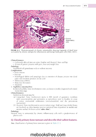
Dead
bone
Chronic
inflammatory
cells
FIGURE 21.3 Photomicrograph of chronic osteomyelitis showing fragments of dead bone
surrounded by chronic nonspecific inflammation and foreign body giant cells (H&E; 2003).
Clinical features:
• Commonly affected sites are spine (lumbar and thoracic), knee and hip
• Almost all patient presents with pain, fever and weight loss
Pathology:
Epithelioid cell granulomas with or without necrosis
Complications:
• Psoas abscess
• Fracture
• Neurological deficit and paraplegia due to extension of disease process into dural
space with resultant pressure on the cord
• Tuberculous arthritis
• Sinus tract formation
• Ankylosis
3. Syphilitic osteomyelitis:
In skeletal syphilis, bone involvement is rare, as disease is readily diagnosed and treated
before this stage.
Clinical features:
Skeletal involvement may be
(a) Congenital: Bone involvement starts in fifth month of gestation; manifests
with osteochondritis and periostitis as spirochetes tend to localize in areas
of active enchondral ossification (osteochondritis) and the periosteum
(periostitis).
(b) Acquired: Bone involvement is seen in tertiary stage. Skull and long tubular bones
are involved, eg, tibia (massive reactive periosteal bone deposition on medial and
anterior surface of tibia is called ‘saber shin’).
Pathology:
Necrotic bone is surrounded by chronic inflammatory cells with a predominance of
plasma cells
Q. Classify primary bone tumours and describe their salient features.
Ans. Classification of primary bone tumours is given in Table 21.1.
mebooksfree.com