
Page 592 - Concise Pathology for Exam Preparation ( PDFDrive )
P. 592
21 Musculoskeletal System 577
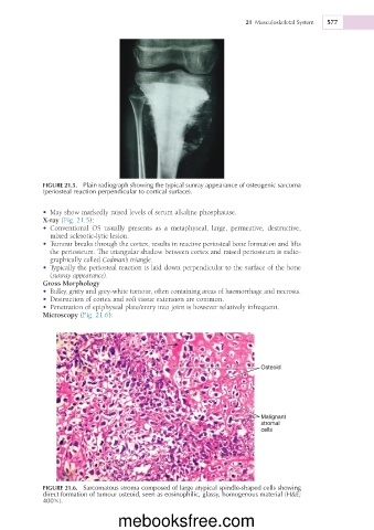
FIGURE 21.5. Plain radiograph showing the typical sunray appearance of osteogenic sarcoma
(periosteal reaction perpendicular to cortical surface).
• May show markedly raised levels of serum alkaline phosphatase.
X-ray (Fig. 21.5):
• Conventional OS usually presents as a metaphyseal, large, permeative, destructive,
mixed sclerotic-lytic lesion.
• Tumour breaks through the cortex, results in reactive periosteal bone formation and lifts
the periosteum. The triangular shadow between cortex and raised periosteum is radio-
graphically called Codman’s triangle.
• Typically the periosteal reaction is laid down perpendicular to the surface of the bone
(sunray appearance).
Gross Morphology
• Bulky, gritty and grey-white tumour, often containing areas of haemorrhage and necrosis.
• Destruction of cortex and soft tissue extension are common.
• Penetration of epiphyseal plate/entry into joint is however relatively infrequent.
Microscopy (Fig. 21.6):
Osteoid
Malignant
stromal
cells
FIGURE 21.6. Sarcomatous stroma composed of large atypical spindle-shaped cells showing
direct formation of tumour osteoid, seen as eosinophilic, glassy, homogenous material (H&E;
4003).
mebooksfree.com