
Page 594 - Concise Pathology for Exam Preparation ( PDFDrive )
P. 594
21 Musculoskeletal System 579
Skeletal Distribution
Metaphysis of lower femur, upper tibia and upper humerus.
Clinical Features
• Solitary osteochondromas are diagnosed in later life as compared to multiple osteochon-
dromas which usually manifest in childhood itself.
• Osteochondromas are mostly asymptomatic but may present with pain and deformity.
They sometimes interfere with the functioning of regional tendons and blood vessels.
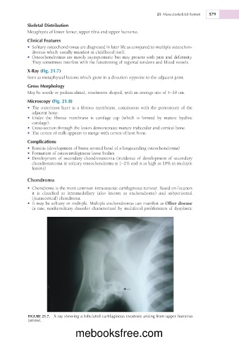
X-Ray (Fig. 21.7)
Seen as metaphyseal lesions which grow in a direction opposite to the adjacent joint.
Gross Morphology
May be sessile or pedunculated, mushroom shaped, with an average size of 4–10 cm.
Microscopy (Fig. 21.8)
• The outermost layer is a fibrous membrane, continuous with the periosteum of the
adjacent bone.
• Under the fibrous membrane is cartilage cap (which is formed by mature hyaline
cartilage).
• Cross-section through the lesion demonstrates mature trabecular and cortical bone.
• The cortex of stalk appears to merge with cortex of host bone.
Complications
• Bursitis (development of bursa around head of a longstanding osteochondroma)
• Formation of osteocartilaginous loose bodies
• Development of secondary chondrosarcoma (incidence of development of secondary
chondrosarcoma in solitary osteochondroma is 1–2% and is as high as 10% in multiple
lesions)
Chondroma
• Chondroma is the most common intraosseous cartilaginous tumour. Based on location
it is classified as intramedullary (also known as enchondroma) and subperiosteal
(juxtacortical) chondroma.
• It may be solitary or multiple. Multiple enchondromas can manifest as Ollier disease
(a rare, nonhereditary disorder characterized by multifocal proliferation of dysplastic
FIGURE 21.7. X-ray showing a lobulated cartilaginous exostosis arising from upper humerus
(arrow).
mebooksfree.com