
Page 596 - Concise Pathology for Exam Preparation ( PDFDrive )
P. 596
21 Musculoskeletal System 581
Lobules of
hyaline
cartilage
Calcification
FIGURE 21.9. Section from an enchondroma showing well-circumscribed nodules of hyaline
cartilage with cytologically benign chondrocytes. The centre of the nodule shows calcification
(H&E; 1003).
X-Ray
Shows a well-defined lytic lesion surrounded by sclerosis. Spotty calcification is common.
Cysts are present about 20% of the time and both MRI and CT can define fluid levels.
Gross Morphology
On gross examination, chondroblastoma has a lobulated, round form and is made up of
friable, soft, greyish-pink tissue that may be gritty.
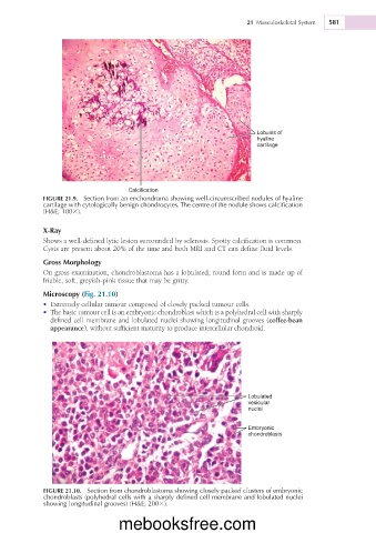
Microscopy (Fig. 21.10)
• Extremely cellular tumour composed of closely packed tumour cells.
• The basic tumour cell is an embryonic chondroblast which is a polyhedral cell with sharply
defined cell membrane and lobulated nuclei showing longitudinal grooves (coffee-bean
appearance), without sufficient maturity to produce intercellular chondroid.
Lobulated
vesicular
nuclei
Embryonic
chondroblasts
FIGURE 21.10. Section from chondroblastoma showing closely packed clusters of embryonic
chondroblasts (polyhedral cells with a sharply defined cell membrane and lobulated nuclei
showing longitudinal grooves) (H&E; 2003).
mebooksfree.com