
Page 598 - Concise Pathology for Exam Preparation ( PDFDrive )
P. 598
21 Musculoskeletal System 583
(b) Secondary chondrosarcoma: Arises from benign cartilage defects such as
osteochondroma or enchondroma
2. Based on topography:
(a) Conventional intramedullary: Arises from the medullary cavity of long bones,
pelvis, costochondral junction of ribs and shoulders and presents as a lytic lesion
with blotchy calcification.
(b) Juxtacortical (peripheral): Arises in the shaft of a long bone.
3. Based on morphology:
(a) Conventional (which is further subtyped as hyaline or myxoid)
(b) Clear cell
(c) Dedifferentiated
(d) Mesenchymal
Gross Morphology
Grey-white, lobulated, bulky, translucent masses with a gelatinous consistency. Erosion/
destruction of cortex is frequently seen. Calcification and ossification are not uncommon.
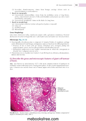
Microscopy (Fig. 21.12)
• Histologically, chondrosarcoma is composed of invasive lobules of anaplastic cartilage
and is differentiated from benign cartilaginous tumours based on the following features:
• Presence of two or more cells per lacuna, binucleate cells, enlarged, plump and
hyperchromatic nuclei, nuclear pleomorphism and abundant mitoses.
• Enchondral ossification is seen (unlike osteosarcoma in which the osteogenesis is
directly from malignant stromal cells).
• Chondrosarcoma is classified into Grades I, II and III, based on cellularity, pleomorphism,
mitoses and necrosis.
Q. Describe the gross and microscopic features of giant cell tumour
of bone.
Ans. Also known as osteoclastoma, GCT is the most common tumour of epiphyses in
skeletally mature individuals with closed growth plates. It often shows metaphyseal exten-
sion. Common sites include lower end of femur, upper end of tibia and lower end of ra-
dius.
Malignant
cartilage with
pleomorphic nuclei
FIGURE 21.12. Section from a chondrosarcoma showing cartilaginous lobules composed of
atypical chondrocytes (H&E; 2003).
mebooksfree.com