
Page 595 - Concise Pathology for Exam Preparation ( PDFDrive )
P. 595
580 SECTION II Diseases of Organ Systems
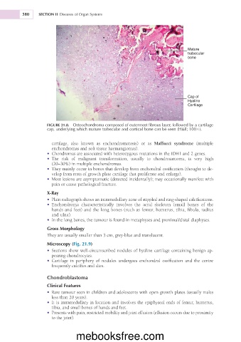
Mature
trabecular
bone
Cap of
Hyaline
Cartilage
FIGURE 21.8. Osteochondroma composed of outermost fibrous layer, followed by a cartilage
cap, underlying which mature trabecular and cortical bone can be seen (H&E; 1003).
cartilage, also known as enchondromatosis) or as Maffucci syndrome (multiple
enchondromas and soft tissue haemangiomas).
• Chondromas are associated with heterozygous mutations in the IDH1 and 2 genes.
• The risk of malignant transformation, usually to chondrosarcoma, is very high
(20–30%) in multiple enchondromas.
• They mainly occur in bones that develop from enchondral ossification (thought to de-
velop from rests of growth plate cartilage that proliferate and enlarge).
• Most lesions are asymptomatic (detected incidentally); may occasionally manifest with
pain or cause pathological fracture.
X-Ray
• Plain radiograph shows an intramedullary zone of stippled and ring-shaped calcifications.
• Enchondroma characteristically involves the acral skeleton (small bones of the
hands and feet) and the long bones (such as femur, humerus, tibia, fibula, radius
and ulna).
• In the long bones, the tumour is found in metaphyses and proximal/distal diaphyses.
Gross Morphology
They are usually smaller than 3 cm, grey-blue and translucent.
Microscopy (Fig. 21.9)
• Sections show well-circumscribed nodules of hyaline cartilage containing benign ap-
pearing chondrocytes.
• Cartilage in periphery of nodules undergoes enchondral ossification and the centre
frequently calcifies and dies.
Chondroblastoma
Clinical Features
• Rare tumour seen in children and adolescents with open growth plates (usually males
less than 20 years).
• It is intramedullary in location and involves the epiphyseal ends of femur, humerus,
tibia, and small bones of hands and feet.
• Presents with pain, restricted mobility and joint effusion (effusion occurs due to proximity
to the joint).
mebooksfree.com