
Page 599 - Concise Pathology for Exam Preparation ( PDFDrive )
P. 599
584 SECTION II Diseases of Organ Systems
Cortical thinning
Lytic expansile
epiphyseal lesion
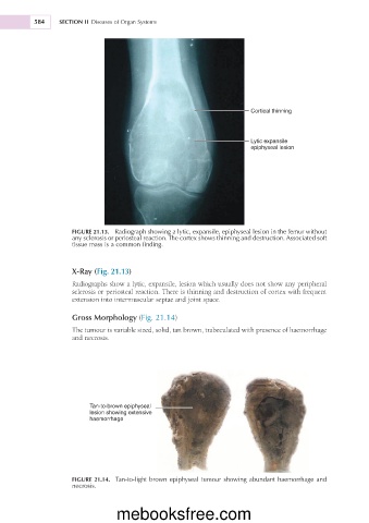
FIGURE 21.13. Radiograph showing a lytic, expansile, epiphyseal lesion in the femur without
any sclerosis or periosteal reaction. The cortex shows thinning and destruction. Associated soft
tissue mass is a common finding.
X-Ray (Fig. 21.13)
Radiographs show a lytic, expansile, lesion which usually does not show any peripheral
sclerosis or periosteal reaction. There is thinning and destruction of cortex with frequent
extension into intermuscular septae and joint space.
Gross Morphology (Fig. 21.14)
The tumour is variable sized, solid, tan brown, trabeculated with presence of haemorrhage
and necrosis.
Tan-to-brown epiphyseal
lesion showing extensive
haemorrhage
FIGURE 21.14. Tan-to-light brown epiphyseal tumour showing abundant haemorrhage and
necrosis.
mebooksfree.com