
Page 597 - Concise Pathology for Exam Preparation ( PDFDrive )
P. 597
582 SECTION II Diseases of Organ Systems
• Mitoses and necrosis are frequent; scattered osteoclastic giant cells may also be seen.
• Scant amount of lace-like hyaline matrix may be laid down, which calcifies to produce
a characteristic chicken-wire calcification.
Chondromyxoid Fibroma (CMF)
Clinical Features
Affects young adults and presents with localized dull aching pain and swelling in the
affected region.
X-Ray
Large, lobulated, sharply defined, eccentric, lytic, metaphyseal lesion surrounded by a rim
of sclerosis.
Gross Morphology
Average size is 3–8 cm; cut surface appears solid, glistening and tan-grey.
Microscopy (Fig. 21.11)
• Prominent features of CMF are the zonal architecture and lobular pattern. Hypocellular
lobules of poorly formed hyaline cartilage and myxoid tissue are separated by fibrous septae.
• The chondrocytes in myxoid areas are plump-to-spindled in shape and have indistinct
cell borders.
• Varying degree of cytological atypia is common along with small foci of calcification.
Chondrosarcoma
It is a malignant mesenchymal tumour that produces cartilaginous matrix. There are sev-
eral subtypes of chondrosarcoma, which vary in terms of location, appearance, treatment
and prognosis.
Classification
1. Based on pre-existing pathology:
(a) Primary chondrosarcoma: Relatively uncommon; arises centrally in the bone, and
is found in children
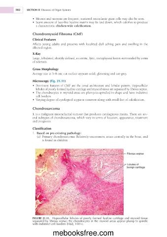
Fibrous septae
Lobules of
benign cartilage
FIGURE 21.11. Hypocellular lobules of poorly formed hyaline cartilage and myxoid tissue
separated by fibrous septae; the chondrocytes in the myxoid areas appear plump to spindle
with indistinct cell borders (H&E; 1003).
mebooksfree.com