
Page 600 - Concise Pathology for Exam Preparation ( PDFDrive )
P. 600
21 Musculoskeletal System 585
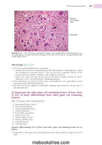
Stromal
mononuclear
cells
Giant cells
FIGURE 21.15. GCT of bone composed of uniform oval mononuclear cells that grow in a
syncytial pattern (stromal element) and giant cells with 20–30 nuclei arranged towards the
centre (H&E; 2003).
Microscopy (Fig. 21.15)
• GCT has two histopathological components:
• Stromal cells: Uniform oval mononuclear cells with indistinct cell membrane, which
are arranged in a syncytial pattern. They are the basic neoplastic element of the
tumour and their number correlates with its clinical evolution.
• Giant cells: Large cells with 20–30 (up to 100) nuclei arranged towards the centre
(believed to form via RANK/RANKL signalling pathway).
• Focal deposition of osteoid or bone may occasionally be seen, especially in cases
presenting with pathological fracture.
• All GCTs should be regarded as potentially malignant (approximately 4% give rise to
distant metastasis).
Q. Enumerate the other giant cell containing lesions of bone. How
is GCT of bone differentiated from other giant cell containing
lesions?
Ans. Other giant cells containing lesions:
1. Metaphyseal fibrous defect
2. Nonossifying fibroma
3. Chondromyxoid fibroma
4. Chondroblastoma
5. Eosinophilic granuloma
6. Solitary bone cyst
7. Osteitis fibrosa cystica
8. Aneurysmal bone cyst
9. Osteoid osteoma
10. Osteoblastoma
Features differentiating GCT of bone from other giant cell-containing lesions are as
follows:
• Giant cells in other giant cell-containing lesions have fewer nuclei as compared to GCT
bone.
mebooksfree.com