
Page 1266 - Hematology_ Basic Principles and Practice ( PDFDrive )
P. 1266
1112 Part VII Hematologic Malignancies
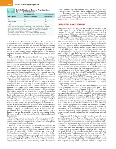
TABLE Risk Stratification in Essential Thrombocythemia plaques, and unexplained dermatoses. Rarely, vascular changes result
69.2 Based on Thrombotic Risk a in microcirculatory flow abnormalities, leading to a vasculitis result-
ing in classic purpura that may progress to skin necrosis. This should
Age >60 Years or Cardiovascular be distinguished from leg ulcers that occur in patients being treated
Risk Category History of Thrombosis Risk Factors with hydroxyurea. Occasionally, patients also develop pyoderma
Low No No gangrenosum or Sweet syndrome.
Intermediate No Yes
High Yes Yes LABORATORY MANIFESTATIONS
a Cardiovascular risk factors: hypertension, hypercholesterolemia, diabetes,
smoking, and congestive heart failure. Extreme thrombocytosis (platelet count The hallmark of ET is a sustained and unexplained elevation of the
>1500 × 10 /L) is a risk factor for bleeding. Its role as a risk factor for platelet count (≥450 × 10 /L). Accompanying leukocytosis is a
9
9
thrombosis in essential thrombocythemia is uncertain.
Data from Finazzi G, Barbui T: Risk-adapted therapy in essential common finding. A leukoerythroblastic blood picture, as well as
thrombocythemia and polycythemia vera. Blood Rev 19:243, 2005. teardrop-shaped RBCs are not features of ET but are suggestive of
3
an early form of MF. Mild eosinophilia (>400/mm ) and basophilia
3
(>100/mm ) have been reported in more than one-third of patients.
The most common morphologic abnormalities are variations in
A meta-analysis has revealed that the JAK2V617 mutation is RBC size and shape and the presence of megathrombocytes (Fig.
associated with a twofold higher risk of developing either a venous 69.3). The BM is usually normocellular or slightly hypercellular
or arterial thrombosis but does not influence the risk of suffering without a significant increase in granulopoiesis or erythropoiesis.
from a hemorrhagic event. Regardless, it is conceivable that the sig- Increased numbers of enlarged megakaryocytes with hyperlobulated
nificantly more advanced age and elevated hematocrit and leukocyte or deeply folded nuclei that cluster in small groups along sinuses are
levels in mutation-positive patients might contribute to the apparent the hallmarks of ET (see Fig. 69.4). Reticulin fibrosis is not signifi-
association between JAK2V617F and thrombosis reported in some cantly evident. A great deal of controversy currently surrounds dis-
studies. tinguishing “true ET” from an early prefibrotic form of MF (prePMF)
Patients with ET who are older than 60 years of age who have in which the BM is characteristically hypercellular with pronounced
had a prior thrombotic event have a greater risk of developing addi- proliferation of granulocytes and reduced erythroid precursors. The
tional thrombotic events (Table 69.2). By contrast, the incidence of megakaryocytes are increased in number but are loosely clustered or
thrombotic and hemorrhagic complications in asymptomatic patients located along the endosteal bone surface. The megakaryocytes contain
with ET who are younger than 60 years of age who have platelet hyperchromatic, hypolobulated bulbous, or irregularly folded nuclei
9
counts of less than 1500 × 10 /L has been shown to be comparable with an abnormal nuclear-to-cytoplasmic ratio. The histopathologic
to a normal control population. Gender, hypertension, diabetes mel- criteria for this form of prePMF have been combined with clinical
litus, hypercholesterolemia, and smoking have been shown to be criteria (minor criteria) by the World Health Organization (WHO).
independent risk factors for developing arterial thrombotic complica- The 2016 criteria for the diagnosis of ET and preMF are particularly
tions in ET (see Table 69.2). Screening of patients for other acquired useful (Table 69.3). These new criteria are heavily dependent on
and inherited thrombophilic states may identify patients at an even mutational analyses. The JAK2V617F mutation occurs in 50–60%
higher risk for both arterial and venous thrombotic events. of ET patients, while recurrent mutations in CALR mutations occur
Pregnancy is not contraindicated in patients with ET. The outcome in 25% of patients and 3–5% have MPL mutations. Approximately
of pregnancy in patients with ET has been the subject of intense 25% of patients harbor mutated CALR as either a type I (52-bp del)
investigation. The rate of having a successful pregnancy is 61% or type II (5-bp insertion).
compared with an 85–90% rate in normal women. The rate of The patients with ET who lack such driver mutations are said to
spontaneous abortions ranges from 39–44% compared with the be “triple negative” (Table 69.3). Based on the 2016 WHO Diag-
miscarriage rate of 10–15% in normal pregnancies. Placental infarc- nostic Criteria, the presence of modest BM reticulin fibrosis does
tion is often responsible for intrauterine fetal growth retardation not exclude the diagnosis of ET. Although select hematopathologists
(5%). Abruptio placenta has been reported in 3.6% of cases, a rate can reproducibly distinguish ET from prePMF, it remains uncertain
that is higher than that observed in the general population (1%). whether this distinction is broadly applicable. In 70–80% of ET
Major thrombotic episodes occur in 3% of these pregnancies while patients, iron stores were present in the BM, albeit at reduced levels.
major bleeding episodes occur in 2% of cases. These rates are higher Almost all patients have normal serum ferritin levels. The absence
than that observed in the overall population of pregnant women. of iron stores in 30% of patients may merely be an epiphenomenon
Baseline platelet count is not predictive of pregnancy outcome. ET of a chronic MPN and not truly reflective of an iron deficiency
patients with the JAK2V617F mutation have been reported to be at state. Platelet aggregation study results are frequently abnormal,
a higher risk of developing complications with pregnancy. In the most often demonstrate an impaired aggregation response to
postpartum period, the platelet counts return to their earlier levels, epinephrine, ADP, and collagen but not to arachidonic acid and
and rebound thrombocytosis may occur in some patients. This is ristocetin. Spontaneous platelet aggregation has been reported to
thought to increase the probability of vascular complications during occur frequently in such patients, but this has not been a universal
this period to a level similar to that observed in other conditions of finding.
thrombophilia. Approximately 25% of patients with ET have been reported to have
In the large majority of cases, the fetal losses in pregnant ET elevated uric acid levels at diagnosis. The average value of the serum
women occur during the first trimester. A previous history of spon- potassium at diagnosis is usually within the normal range, although
taneous abortion may be the greatest risk factor for the development 23% of patients have been reported to have pseudohyperkalemia,
of subsequent spontaneous abortions. caused by the degranulation of platelets when in vitro clotting releases
Physical examination findings are relatively unremarkable in potassium. Both excessive numbers of RBCs and leukocytes can also
patients with ET. Most patients are not severely ill at diagnosis, with be associated with these phenomena. The pseudohyperkalemia can
a median Karnofsky score of 90% being reported in one series. be documented by measuring plasma instead of serum potassium
Splenomegaly is detectable in 40–50% of patients, and approximately levels and the lack of electrocardiographic findings associated with
20% have hepatomegaly. During the course of the disorder, a further true hyperkalemia. Pseudohypoxemia has also been observed in ET
increase in the degree of hepatosplenomegaly may be observed in patients with extreme degrees of thrombocytosis. Acquired von Wil-
patients who are developing post-ET MF. Recently, skin manifesta- lebrand syndrome is associated almost uniformly with a platelet count
9
tions of the MPN have been reported to be relatively common, greater than 1500 × 10 /L, a prolonged bleeding time, normal factor
ranging from paraneoplastic lesions, including vascular, neutrophilic VIII coagulant activity, and a normal von Willebrand antigen level