
Page 1268 - Hematology_ Basic Principles and Practice ( PDFDrive )
P. 1268
1114 Part VII Hematologic Malignancies
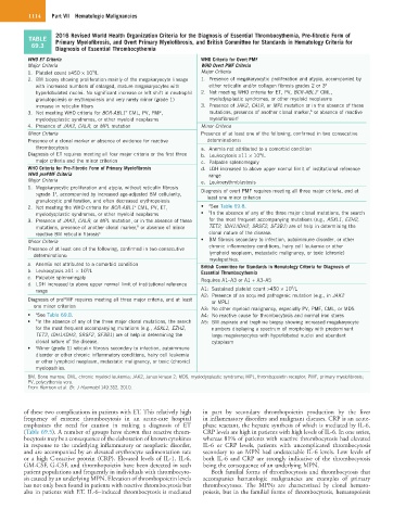
2016 Revised World Health Organization Criteria for the Diagnosis of Essential Thrombocythemia, Pre-fibrotic Form of
TABLE Primary Myelofibrosis, and Overt Primary Myelofibrosis, and British Committee for Standards in Hematology Criteria for
69.3 Diagnosis of Essential Thrombocythemia
WHO ET Criteria WHO Criteria for Overt PMF
Major Criteria WHO Overt PMF Criteria
9
1. Platelet count ≥450 × 10 /L Major Criteria
2. BM biopsy showing proliferation mainly of the megakaryocyte lineage 1. Presence of megakaryocytic proliferation and atypia, accompanied by
with increased numbers of enlarged, mature megakaryocytes with either reticulin and/or collagen fibrosis grades 2 or 3 a
+
hyperlobulated nuclei. No significant increase or left shift in neutrophil 2. Not meeting WHO criteria for ET, PV, BCR-ABL1 CML,
granulopoiesis or erythropoiesis and very rarely minor (grade 1) myelodysplastic syndromes, or other myeloid neoplasms
increase in reticulin fibers 3. Presence of JAK2, CALR, or MPL mutation or in the absence of these
b
3. Not meeting WHO criteria for BCR-ABL1 CML, PV, PMF, mutations, presence of another clonal marker, or absence of reactive
+
myelodysplastic syndromes, or other myeloid neoplasms myelofibrosis c
4. Presence of JAK2, CALR, or MPL mutation Minor Criteria
Minor Criteria Presence of at least one of the following, confirmed in two consecutive
Presence of a clonal marker or absence of evidence for reactive determinations:
thrombocytosis a. Anemia not attributed to a comorbid condition
Diagnosis of ET requires meeting all four major criteria or the first three b. Leukocytosis ≥11 × 10 /L
9
major criteria and the minor criterion c. Palpable splenomegaly
WHO Criteria for Pre-Fibrotic Form of Primary Myelofibrosis d. LDH increased to above upper normal limit of institutional reference
WHO prePMF Criteria range
Major Criteria e. Leukoerythroblastosis
1. Megakaryocytic proliferation and atypia, without reticulin fibrosis Diagnosis of overt PMF requires meeting all three major criteria, and at
>grade 1 , accompanied by increased age-adjusted BM cellularity, least one minor criterion
a
granulocytic proliferation, and often decreased erythropoiesis
2. Not meeting the WHO criteria for BCR-ABL1 CML, PV, ET, • a See Table 69.8.
+
myelodysplastic syndromes, or other myeloid neoplasms • b In the absence of any of the three major clonal mutations, the search
3. Presence of JAK2, CALR, or MPL mutation, or in the absence of these for the most frequent accompanying mutations (e.g., ASXL1, EZH2,
b
mutations, presence of another clonal marker, or absence of minor TET2, IDH1/IDH2, SRSF2, SF3B1) are of help in determining the
reactive BM reticulin fibrosis c clonal nature of the disease.
Minor Criteria • BM fibrosis secondary to infection, autoimmune disorder, or other
Presence of at least one of the following, confirmed in two consecutive chronic inflammatory conditions, hairy cell leukemia or other
determinations: lymphoid neoplasm, metastatic malignancy, or toxic (chronic)
myelopathies.
a. Anemia not attributed to a comorbid condition British Committee for Standards in Hematology Criteria for Diagnosis of
b. Leukocytosis ≥11 × 10 /L Essential Thrombocythemia
9
c. Palpable splenomegaly Requires A1–A3 or A1 + A3–A5
d. LDH increased to above upper normal limit of institutional reference 9
range A1: Sustained platelet count >450 × 10 /L
Diagnosis of prePMF requires meeting all three major criteria, and at least A2: Presence of an acquired pathogenic mutation (e.g., in JAK2
or MPL)
one minor criterion A3: No other myeloid malignancy, especially PV, PMF, CML, or MDS
• a See Table 69.8. A4: No reactive cause for thrombocytosis and normal iron stores
• b In the absence of any of the three major clonal mutations, the search A5: BM aspirate and trephine biopsy showing increased megakaryocyte
for the most frequent accompanying mutations (e.g., ASXL1, EZH2, numbers displaying a spectrum of morphology with predominant
TET2, IDH1/IDH2, SRSF2, SF3B1) are of help in determining the large megakaryocytes with hyperlobated nuclei and abundant
clonal nature of the disease. cytoplasm
• c Minor (grade 1) reticulin fibrosis secondary to infection, autoimmune
disorder or other chronic inflammatory conditions, hairy cell leukemia
or other lymphoid neoplasm, metastatic malignancy, or toxic (chronic)
myelopathies.
BM, Bone marrow; CML, chronic myeloid leukemia; JAK2, Janus kinase 2; MDS, myelodysplastic syndrome; MPL, thrombopoietin receptor; PMF, primary myelofibrosis;
PV, polycythemia vera.
From Harrison et al: Br. J Haemotol 149:352, 2010.
of these two complications in patients with ET. This relatively high in part by secondary thrombopoietin production by the liver
frequency of extreme thrombocytosis in an acute-care hospital in inflammatory disorders and malignant diseases. CRP is an acute-
emphasizes the need for caution in making a diagnosis of ET phase reactant, the hepatic synthesis of which is mediated by IL-6.
(Table 69.5). A number of groups have shown that reactive throm- CRP levels are high in patients with high levels of IL-6. In one series,
bocytosis may be a consequence of the elaboration of known cytokines whereas 81% of patients with reactive thrombocytosis had elevated
in response to the underlying inflammatory or neoplastic disorder, IL-6 or CRP levels, patients with uncomplicated thrombocytosis
and are accompanied by an elevated erythrocyte sedimentation rate secondary to an MPN had undetectable IL-6 levels. Low levels of
or a high C-reactive protein (CRP). Elevated levels of IL-1, IL-6, both IL-6 and CRP are strongly indicative of the thrombocytosis
GM-CSF, G-CSF, and thrombopoietin have been detected in such being the consequence of an underlying MPN.
patient populations and frequently in individuals with thrombocyto- Both familial forms of thrombocytosis and thrombocytosis that
sis caused by an underlying MPN. Elevation of thrombopoietin levels accompanies hematologic malignancies are examples of primary
has not only been found in patients with reactive thrombocytosis but thrombocytoses. The MPNs are characterized by clonal hemato-
also in patients with ET. IL-6–induced thrombocytosis is mediated poiesis, but in the familial forms of thrombocytosis, hematopoiesis