
Page 1334 - Hematology_ Basic Principles and Practice ( PDFDrive )
P. 1334
1180 Part VII Hematologic Malignancies
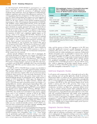
the t(8;21)(q22;q22) RUNX1/RUNX1T rearrangement is a well- Clinicopathologic Features of Eosinophilia-Associated
known association. In cases of KIT D816V-positive SM, several TABLE FIP1L1-PDGFRA–Rearranged Myeloid Neoplasms
reports have now detailed the coexistence of additional myeloid 72.9 Versus KIT D816V-Positive Systemic Mastocytosis
neoplasm-related mutations, which are implicated in differentiation,
epigenetic regulation, and control of the spliceosome machinery. The FIP1L1-PDGFRA–
common themes that have emerged are: (1) ISM tends to be more a Features Rearranged KIT D816V–Positive
pure KIT D816V-driven disease with absence of, or low frequency of Gender Overwhelmingly male Less gender skewing
additional (known) molecular abnormalities; (2) TET2, SRSF2, and
ASXL1 are the most common of the myeloid neoplasm-associated Bone marrow mast Loose clusters/ Dense aggregates
mutated genes (~20%–35% of patients), but mutations in DNMT3A, cell aggregates interstitial
CBL, K/NRAS, UA2F1, ZRSF2 EZH2, RUNX1, ETV6, and SETBP1 AEC/tryptase ratio >100 ≤100
have also been identified; and (3) the presence of one or more of these Treatment Imatinib sensitive Imatinib-resistant;
genes mutations are usually associated with more advanced disease second-generation TKIs
subtypes, particularly SM-AHN. Schwaab et al found that the pres- (e.g., midostaurin)
ence of one or more of these mutations in addition to KIT D816V
22
was associated with inferior survival, and a separate analysis found Symptom profile Cardiac/pulmonary Gastrointestinal/urticaria
that mutation of ASXL1 had a significant negative impact on OS pigmentosa/anaphylaxis
among a cohort of 62 patients with SM-AHN. Mutations in the Vitamin B 12 level Elevated Often normal
ethanolamine kinase gene ETNK1 were recently found to be enriched AEC, Absolute eosinophil count; TKI, tyrosine kinase inhibitor.
in SM patients with eosinophilia, but are commonly present with Modified from Maric I, Robyn J, Metcalfe DD, et al: KIT D816V-associated
KIT D816V or other myeloid disease-associated mutations (Dr. systemic mastocytosis with eosinophilia and FIP1L1/PDGFRA-associated
Andreas Reiter, personal communication). Mutations in ETNK1 chronic eosinophilic leukemia are distinct entities. J Allergy Clin Immunol
120(3):680-687, 2007.
were also identified in ~9% of patients with atypical CML and in
<5% of patients with CMML. The basis for the restriction of ETNK1
mutations to these diseases and how they contribute to their patho-
genesis is unknown. In one patient with MCL, exome sequencing value, and the presence of dense MC aggregates in the BM were
revealed an imatinib-resistant KIT V654A mutation as well as a statistically significantly increased or more frequently represented
mutation in the β-chain of FcεRI, but the functional consequences compared with patients with the FIP1L1-PDGFRA gene fusion.
of this variant were not explored. Conversely, male sex, cardiac and pulmonary symptoms, median peak
Special research techniques have been used to interrogate the absolute eosinophil count, the eosinophil to tryptase ratio, and ele-
cell-specific distribution of mutations. For example, KIT D816V has vated serum B 12 levels were higher or more common in the group
been demonstrated in both MCs and the AHN component (e.g., with FIP1L1-PDGFRA. A scoring system incorporating these clinical
monocytes in CMML) by cell laser microdissection; TET2 and KIT and laboratory parameters could reliably predict whether patients
D816V have been found together in flow-sorted MCs; the SRSF2 with peripheral eosinophilia and increased marrow MC burden
P95 hotspot mutation has been uncovered in both MCs and mono- carried the FIP1L1-PDGFRA gene fusion or the KIT D816V muta-
23
cytes from a case of SM-AHN; and finally, t(8;21) has been identified tion, which is important for guiding targeted therapy options.
by FISH in the BM MCs and the leukemic clone of a patient with Treatment of FIP1L1-PDGFRA–positive myeloid neoplasms associ-
SM and AML. ated with eosinophilia is discussed in Chapter 71.
In 2003 the FIP1L1-PDGFRA fusion oncogene was identified in
patients with a diagnosis of idiopathic hypereosinophilic syndrome.
This molecular abnormality is not visible by standard chromosome Additional Diagnostic Studies
analysis and its detection requires either use of reverse-transcriptase
polymerase chain reaction or, more commonly, fluorescence in situ In all patients with symptomatic SM, a thorough work-up for aller-
hybridization (FISH). Because this genetic lesion results from an gies, including total and specific IgE levels, should be performed.
interstitial 800-kb deletion on chromosome 4q12, which removes a Biochemical studies to evaluate for MC activation/mediator release
segment of DNA involving the CHIC2 gene, the diagnostic test include 24-h urine evaluation of N-methylhistamine, prostaglandin
is referred to as “FISH for the CHIC2 deletion.” Patients with D2, or 11-B-prostaglandin F2. Bleeding diatheses have been reported
FIP1L1-PDGFRA-associated eosinophilia present with features of a in patients with MCAS or SM, and increased heparin levels can be
myeloproliferative disorder: splenomegaly, hypercellular BMs, and found in patients with SM or MC activation. Dual energy x-ray
clinicopathologic characteristics that overlap with SM, including absorptiometry (DEXA) scans should be undertaken to screen for
increased numbers of abnormal-appearing BM MCs, marrow fibrosis, osteoporosis, and plain x-ray films (e.g., metastastic skeletal survey)
and elevated serum tryptase levels. However, although FIP1L1- are used to evaluate osteolytic lesions and/or pathologic fractures.
PDGFRA–positive myeloid neoplasms may exhibit increased numbers MC-related gastrointestinal symptoms such as diarrhea and signs of
of BM MCs, they usually form interstitial or loose clusters, and this malabsorption (e.g., hypoalbuminemia and weight loss) can be evalu-
disease entity is not considered a subtype of SM by the WHO. ated with endoscopy/colonoscopy with biopsy using appropriate
However, in a smaller subset of patients, criteria for SM may be immunohistochemical stains (e.g., CD117, tryptase, CD25, and
fulfilled even if a KIT mutation is not detected. Notably, in almost all CD3 as a control T-cell marker) to highlight abnormal MCs. Ultra-
cases with FIP1L1-PDGFRA–positive myeloid neoplasms, including sonography, CT and/or MRI of the abdomen/pelvis (with or without
those who have SM-CEL, MCs do not exhibit KIT D816V, even if volumetric imaging) are utilized to determine the presence and extent
they express CD25. Although the FIP1L1-PDGFRA genetic abnor- of B and C findings such as hepato/splenomegaly, portal hyperten-
mality was once considered mutually exclusive of KIT D816V, rare sion, ascites, and lymphadenopathy.
cases with both mutations have now been reported, including their
coexistence in tryptase-positive microdissected MCs.
Investigators identified a set of clinical and laboratory features that Diagnostic Decision Making
could reliably distinguish patients with KIT D816V-positive SM with
eosinophilia from individuals with the FIP1L1-PDGFRA–positive In patients presenting with MC activation symptoms or anaphylaxis
23
myeloid neoplasms with eosinophilia (Table 72.9). These criteria without signs of cutaneous involvement, and a normal or elevated
may be particularly useful when molecular testing is not readily serum tryptase level, the diagnostic workup is centered on establish-
available. In the D816V KIT-positive group, gastrointestinal symp- ing whether WHO diagnostic criteria for SM are met (Fig. 72.7). If
toms, urticaria pigmentosa, thrombocytosis, median serum tryptase these criteria are not satisfied, a workup is performed to rule out or